
Download
REVIEW ARTICLE
Microplastics exposure and immunologic response
Marilyn Urrutia-Pereiraa, Guillermo Guidos-Fogelbachb, Herberto José Chong-Netoc*, Dirceu Soléd
aFaculty of Medicine, Federal University of Pampa, Uruguaiana, Brazil
bEscuela Nacional de Medicina y Homeopatia, Instituto Politécnico Nacional, Ciudad de Mexico, Mexico
cDepartment of Pediatrics, Divison of Allergy and Pneumology, Federal University of Paraná, Curitiba, Paraná, Brazil
dDepartment of Pediatrics, Division of Allergy, Clinical Immunology and Rheumatology, Federal University of São Paulo, São Paulo, Brazil
Abstract
Objective: To assess the impact of microplastics (MPs) on human health.
Data Source: The authors conducted a non-systematic review of articles published in English, Portuguese, French, and Spanish in the last decade in the following databases: PubMed, Google Scholar, EMBASE, and SciELO. The keywords used were: microplastics OR nanoplastics OR marine litter OR toxicology OR additives AND human health OR children OR adults.
Data summary: MPs are a group of emerging contaminants that have attracted scientific interest and societal attention in the last decade due to their ubiquitous detection in all environments. Humans can primarily be exposed to MPs and nanoplastics via oral and inhalation routes, but dermal contact cannot be overlooked, especially in young children. The possible toxic effects of plastic particles are due to their potential toxicity, often combined with that of leachable additives and adsorbed contaminants.
Conclusions: Unless the plastic value chain is transformed over the next two decades, the risks to species, marine ecosystems, climate, health, economy, and communities will be unmanageable. However, along with these risks are the unique opportunities to help transition to a more sustainable world.
Key words: Allergy, Epithelia, Microplastics, Nanoplastics
*Corresponding author: Herberto José Chong-Neto, Department of Pediatrics, Divison of Allergy and Pneumology, Federal University of Paraná, Curitiba, Paraná, Brazil. Email address: [email protected]
Received 7 February 2023; Accepted 24 May 2023; Available online 1 September 2023
Copyright: Urrutia-Pereira M, et al.
License: This open access article is licensed under Creative Commons Attribution 4.0 International (CC BY 4.0). http://creativecommons.org/licenses/by/4.0/
Introduction
Plastic is a synthetic material widely used by humans due to its cost-effectiveness, durability, lightweight, and easy to manufacture. It occupies a sizeable place in human or public society, being omnipresent in everyday life, and thus it is important to determine the their potential risks and impacts on the global environment and human health.1
Global plastic production has risen from two million tons in 1950 to 348 million in 2017, making it a USD 522.6-billion industry. This number is estimated to double by 2040, according to a report from the nongovernmental organization, Pew Charitable Trusts.2
The increased concern about the ecological consequences of such materials being in different ecosystems has led to studies involving plastic waste incorporating new concepts, and the term “microplastics” (MPs) was introduced in 2004.3
Although several definitions have been used for MPs, especially for their size, the most used one defines them as particles of synthetic organic polymers of less than 5 mm (National Oceanic and Atmospheric Administration – NOAA).4
The (ISO/TR 21960:2020) standard was published in 2020, entitled “Plastics – Environmental Aspects – State of Knowledge and Methodologies,” where the term “microplastic” was defined as any water-insoluble solid plastic particle with dimensions between 1 μm and 1000 mm. Plastics can be characterized according to their size: as nanoplastics (<100 nm; NPs), MPs (between 100 nm and 5 mm), mesoplastics (between 5 and 20 mm), macroplastics (>20 mm), and megaplastics (>100 mm).5–7
MPs can be classified according to the origin: (i) primary, if they are intentionally released into the environment (used in industry and in personal care products), and (ii) secondary, if they are released indirectly into the environment and suffer progressive degradation due to processes of photo- and thermo-oxidation.8
MPs come in different forms and shapes (such as fibers, fragments, spheres, granules, films, flakes, pellets, and foam). The composition of the original plastic determines the deterioration processes that occur on the surface of the plastic and the time it remains in the environment.9 Some plastics may be chemically harmful, either directly toxic or because they absorb and carry other components, making them hazardous, and they are categorized as follows: physical, chemical, and biological.10 The potential of MPs to cause physical harm to organisms depends on their size and shape.9
Recent studies have found that including morphological attributes when analyzing MP samples can reveal significant differences. Using only the number of particles as a parameter expresses a restricted dimension and ignores the fact that particles of different sizes can cause different effects in ecosystems.11
The following plastic polymers are notably found in MP and NP particles: polyester (polycyclohexylenedimethylene terephthalate [PCT]), polypropylene (PP); polyvinyl chloride (PVC), polystyrene (PS), teflon, nylon 6.6, polyethylene (PE), polyethylene terephthalate (PET), styrene acrylonitrile resin (SAN), and poly(n-butyl methacrylate) (PBMA).12
Internal Environment
MPs are a threat to the marine ecosystems, and these polluting particles have been documented in different environments in recent years.8 Studies that assessed the presence of MPs in indoor environments (air and dust from homes and offices) documented a very high concentration compared to the outdoor environment.13 Internal environments include private residences, workplaces (e.g., offices), buildings, schools, universities, sports halls, and public transportation.8
There are several direct and indirect sources of plastics in indoor environments. These include personal-care and personal-use products, paints, synthetic grass in sports halls, floor coatings, furniture and fibers, and 3-D printers.13
This study defined the concentration of MP in three indoor environments, namely, houses, public transport, and working places, which are representative of urban life.14 The highest mean MP concentrations were found on buses (17.3 ± 2.4 MP/m3), followed by subways (5.8 ± 1.9 MP/m3), residences (4.8 ± 1.6 MP/m3), and workplaces (4.2 ± 1.6 MP/m3). Polyamide, PA (51%), polyester PES (48%), and PP (1%) were the polymers identified and also being the most common ones in personal care products and synthetic textiles.14
In another study, indoor dust samples from residential areas (apartments) had the highest abundance of MPs, followed by offices (896 MPs/g; n = 50), business hotels (843 MPs/g; n = 53), university dormitories (775 MPs/g; n = 48), and university classrooms (209 MPs/g; n = 44).15
Soltani et al. assessed the presence of MPs in Australian homes, which demonstrated that children under 6 months of age faced the highest exposure risk. PE, PS, PA, and polyacrylic (PAC) fibers were found in homes with carpet as the main floor covering and PVC fibers in homes where carpet was absent.16
The average indoor MP concentration in beauty salons was 46 ± 55 MPs/m3. The predominant polymer in indoor air was PAC (27%), followed by rubber (21%) and polyurethane (PU, 13%). Air conditioners, nail treatment, ceiling and flooring with plastic materials, and the number of occupants were the factors affecting indoor MP concentrations.17
Also during washing of clothes, numerous natural or synthetic fibers are released into the waterways (PS, PAC). These fibers are dumped into the domestic wastewater treatment systems and sent to the wastewater treatment plants. However, the amount of MPs significantly impacts the surrounding environments of places with no collection network or sewage treatment.18
Outdoor Environment
MP concentrations in road dust demonstrated a significant relationship with the volume of vehicles, suggesting that traffic is associated with increased concentrations.19 Tire wear particles (TWPs), consisting of styrene butadiene rubber (SBR), are one of the most significant sources of MP worldwide, as are the road-marking paint particles created by the physical abrasion from vehicles and weathering.8
Transport (ambient wind flow), dispersion (local turbulence or disturbance), and deposition (downward air movement) are the various modes of movement of MPs, aided by their size, length, and shape.20
MPs also serve as an ecosystem for organisms called the plastisphere (plastispheres are communities that have evolved to live in human-made plastic environments, including fungi, bacteria, algae, and viruses) and may act as dispersers of these organisms to other environments and lead to global health consequences.11
MPs also interact with pesticides, persistent organic pollutants, and heavy metals and act as vectors for carrying contaminants in different environments21 and transporting microorganisms (including pathogens) by forming a biofilm on the MP surface.22,23 They also significantly increase the transfer efficiency of antibiotic-resistance genes (ARGs).24
Ortega et al. found that MPs can act as carriers of coexisting contaminants in the atmosphere, yielding a better understanding of the dangers and risks of atmospheric and airborne MPs or NPs, their impacts, cotransport capacity, and interaction with the environment.25
Pathways of Exposure
MP pollution in ecosystems has increased as a result of human activities. This has led humankind to suffer from chronic exposure to MPs and NPs through different pathways such as ingestion, inhalation, and dermal contact.26
The amount of MPs an individual is exposed to is another significant point of concern. A systematic review study assessed the average load of MPs and NPs that humans are exposed to through the digestive and respiratory pathways. Table 1 summarizes these results based on the rigor of study selection and validated and comparable quantification methods. Despite this, the amount of MPs and NPs that may lead to the onset of disease in an individual is unknown.26
Table 1 Overall estimate of total human exposure to micro/nanoplastics (MP/NP) through different pathways.26
| Pathways of exposure | Product | Consumption estimated/recommended | Consumption of MP/NP | |||
|---|---|---|---|---|---|---|
| Mean | Daily | Annually | Annual total | |||
| Ingestion | Fruits/vegetables | 400 g/day | 132 749 p/g | 53.09 × 10⁶ | 19.38 × 10⁹ | 2.93 × 10¹⁰ |
| Seafood | 22.41 kg/year | 0.98 p/g | 60.38 | 22.04 × 10³ | ||
| Bottled water | 2 L/day | 13.55 × 10⁶ p/L | 27.10 × 10⁶ | 9.89 × 10⁹ | ||
| Salt | 5 g/day | 142.80 p/kg | 0.71 | 260.61 | ||
| Alcohol | 6.40 L/year | 4.05 p/L | 0.07 | 25.92 | ||
| Inhalation | Air | 8.64 m3/day | 0.68 p/m3 | 5.92 | 2.16 × 10³ | 2.16 × 10³ |
p/g: particles per gram; p/L: particles per liter; p/m3: particle per cubic meter.
Inhalation
As a type of emerging pollutant, MPs are easily ingested by many organisms. Constant exposure to the open air increases the risk of fragmentation and mixing in the atmosphere, enhancing its deleterious action on biological functions due to its small size, high specific surface area, and strong biological penetration capability.27,28
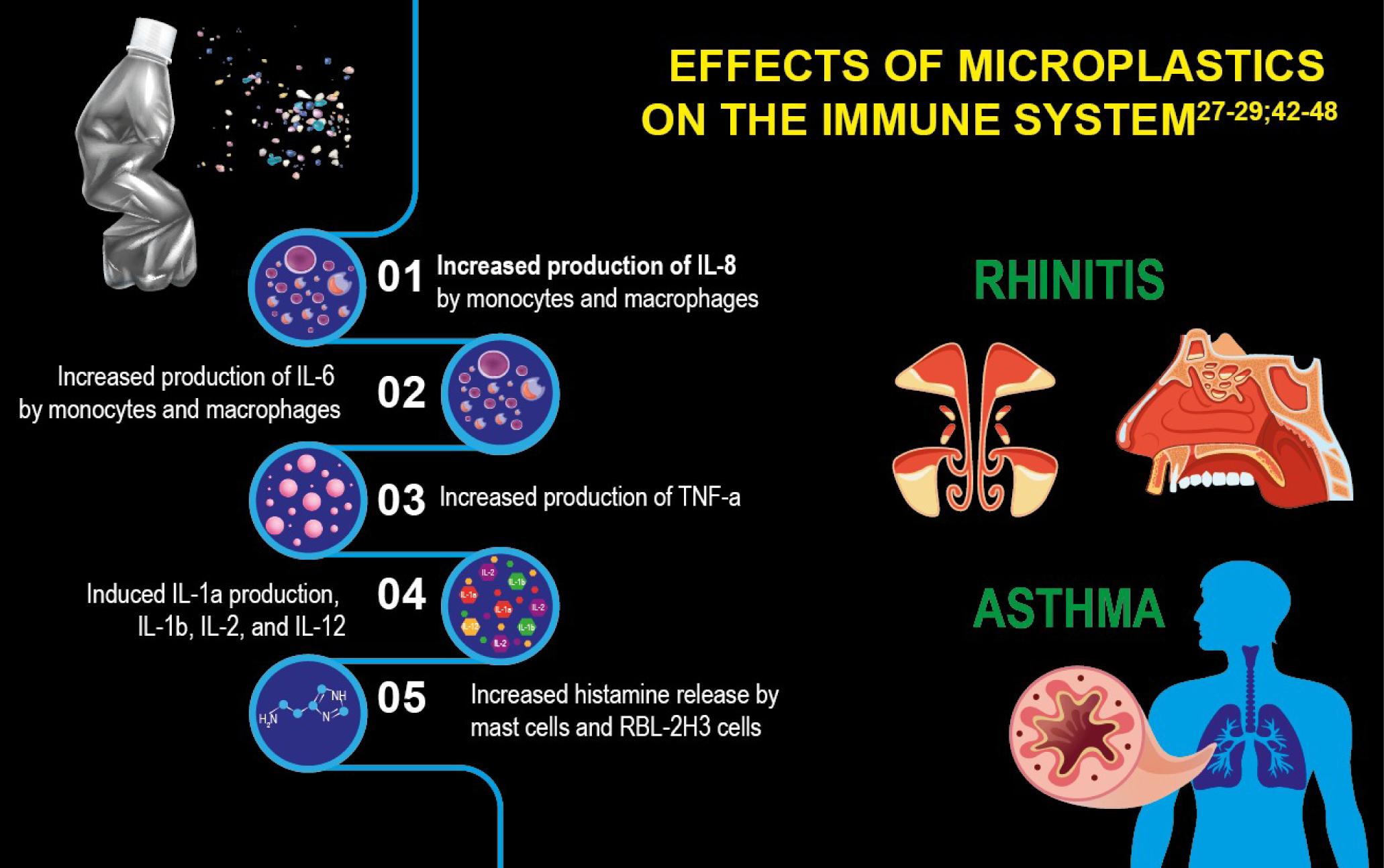
The alveolar surface area of an adult lungs is approximately 150 m2 and has an incredibly thin tissue barrier (<1 μm), making it easier for MP to penetrate and reach the blood capillary system and subsequently disperse throughout the human body.29 Table 2 compiles the effects of MPs on the immune system.
Table 2 Pathways of exposure, effects on immune system, and consequences in cellular and human health.27, 42–58
| Pathways of exposure | Effects on immune system | Cellular and human health* |
|---|---|---|
| Digestive: The main pathway of exposure associated with the ingestion of food contaminated with plastics (water from plastic bottles and/or contaminated with plastics, salt, mollusks, and fish, to name a few). | Increased production of IL-8 by monocytes and macrophages | Digestive system: Cytotoxicity associated with oxidative stress Increased lipid peroxidation Increased reactive oxygen species Increased production of gastric mucus Associated with intestinal microbiota dysbiosis |
| Respiratory: Major sources of exposure come from particles released from car tires, synthetic clothing fibers, and other sources that settle on surfaces such as walls or furniture. | Increased production of IL-6 by monocytes and macrophages | Nervous system: Neurotoxicity secondary to the action of pro-inflammatory cytokines at the neuronal level Some studies suggest a probable association with neurodegenerative diseases |
| Cutaneos: Small particles. Alteration of the epithelial barrier |
Increased production of TNF-α | Respiratory system: Association of autophagy in response to oxidative stress and cytokine-mediated inflammation Some studies suggest a probable association with exacerbating symptoms in chronic respiratory diseases. Alteration of the epithelial barrier is associated with increased prevalence and severity of allergic and inflammatory diseases. |
| Induced IL-1α production, IL-1β, IL-2, and IL-12 | ||
| Increased histamine release by mast cells and RBL-2H3 cells |
*Effects associated with concentration and exposure time.
The epithelial barriers of the skin, respiratory system, and gastrointestinal tract are the first line of physical, chemical, and immunological defense against biological and chemical threats from the surrounding environment. The loss of physical integrity of the barrier due to the degraded proteins in intercellular tight junctions triggers epithelial alarms and the production of cytokines such as interleukin (IL)-25, IL-33, and thymic stromal lymphopoietin, with a consequent increase in epithelium barrier permeability, further facilitating the penetration of allergens, irritants, and microorganisms.30 As a result, consequent changes in the airway microbiota were also observed.
Effects of MPS/NPS on the Respiratory Pathway
Dysfunction of the epithelial barrier, cytotoxicity, synergy with allergens, inflammatory response, and redox imbalance have been the main respiratory health mechanisms identified to be affected by MP or NP exposure.31 When reaching the air–water interface in the alveolus, the MPs or NPs form vesicles that cause biophysical dysfunction of the surfactant, interrupting its structure and mobility and altering the surface tension, and determining its collapse32 to a greater or lesser extent and intensity depending on the type of MP or NP involved33 (Figure 1).
Figure 1 Interactions and possible mechanisms of action of microplastics on the respiratory system. (Adapted from Lu et al.31)
Furthermore, MP exposure alters alveolar structure and airway barrier function. Recent studies have shown that MP can significantly reduce cell viability in a dose-dependent manner, cause major changes in cell morphology, and induce oxidative stress and inflammatory responses, followed by cell death and destruction of the epithelial barrier, which can result in tissue damage and lung disease after prolonged exposure.34–36
The loss of epithelial barrier function resulting from the MP or NP effects facilitates the penetration of allergens, irritants, and microorganisms that allows the onset of an inflammatory process. A typical type 2 immune response in the affected organs favors the development of asthma, rhinitis, or chronic rhinosinusitis.30,37
MPs can also affect nasal microecosystems. High exposure to MPs can increase the abundance of nasal microbiota associated with respiratory tract diseases, such as Klebsiella and Helicobacter, and reduce the abundance of beneficial ones such as bacteroides.4
MPs have raised considerable concerns about the potential risk to children’s health arising from child-specific behaviors, especially in critical periods of neurobehavioral, immune, metabolic, cardiovascular, and other essential body systems, making the early years a critical window to prevent long-lasting damage to health.38
An experimental study with pregnant rats documented the prevalence of NP in their lungs, hearts, and spleens. The same occurred with regard to the placenta and fetal tissues, such as the liver, lungs, heart, kidney, and brain. These facts suggest that the fetoplacental unit is rendered vulnerable to adverse effects on the fetus.39 MP fragments were found in human placentas (fetal side, maternal side, and chorioamniotic membrane) with spherical or irregular shapes and size ranging from 5 to 10 μm.40
Recent research has shown that MP in trophoblastic cytoplasms was associated with decreased cell viability, cell cycle arrest, reduced cell migration and invasion abilities, increased levels of intracellular reactive oxygen species, production of proinflammatory cytokines (TNF-α and IFN-γ) in a dose-dependent manner, and significant changes in the epithelial barrier.38,41 Table 2 summarizes the pathways of exposure, effects on immune system, and consequences of cellular and human health.
Other potential impacts on children’s health could be a result of MP exposure to ambient air in nurseries and schools, generated by various items and activities such as school uniforms, sweatshirts, felt and rubber toys, packaging, paintings and works of art, shoe wear, plastic or laminated furniture, through windows or doors, and the outdoor environment, likely related to location, land use, proximity to traffic and industries, and the degree of ventilation of the building.38,59
An experimental study of mice with asthma exposed to MP documented pulmonary infiltration by inflammatory cells, aggregation of bronchoalveolar macrophages, increased levels of tumor necrosis factor-alpha (TNF-α) in bronchoalveolar lavage, and a worsening of asthma symptoms. Non-asthmatic animals showed an increase in IgG1. Using comparative transcriptomic analysis, the authors documented that MP exposure altered clusters of genes related to immune response, cellular stress response, and programmed cell death, with detrimental outcomes on the mice models.60
Constant exposure to an outdoor environment increased the risk of fragmentation and atmospheric mixing, which facilitated MP availability.28,36 The analysis of human lung tissues obtained during autopsies by Amato-Lourenço et al. demonstrated the presence of PET and PP MPs smaller than 5.5µm in size, and fibers ranging from 8.12 to 16.8 µm.61 The analysis of sputum samples by a few other authors found 21 types of MP smaller than 500 μm, with PU being dominant, followed by PE, PCT, chlorinated PET, and alkyd varnish, mainly related to smoking.62 This was observed on determination of the MP concentration in bronchoalveolar lavage fluid of smokers and nonsmokers, which was significantly higher in smokers.63
A recent study analyzed tumor lesions in patients with or without exposure risk factors, mainly smoking, and documented the presence of microfibers more frequently among exposed patients. The abundance of microfibers in lung tissue gradually accumulated as age increased. These findings reinforce the damaging effects of MPs in cigarette butts.64
Although the cigarette filter has been understood as a barrier that aims to eliminate toxins, thus reducing damage, it is currently known that it does not offer health benefits to smokers. Cigarette filters are composed of more than 15,000 fiber strands which can be put into a microfiber size range (microfiber/MP) or eventually get fragmented into smaller sizes.65
Thus, a new form of cigarette pollution is added to the environment with proven damage to ecosystems and, therefore, to human beings. The correct management of cigarette filters is an unresolved issue that deserves urgent attention and must be addressed by the scientific and social communities as viable source of MPs are capable of harming our planet and its biodiversity.66,67
Digestive
MPs are generated by multiple mechanisms and can be transported through different environmental compartments, reaching the food web and, ultimately, the human body.68
Potential risks to human health from unintentional ingestion of MP and NP are an emerging concern.69 The finding of MP in human feces has proved that these particles are indeed ingested and can pass through the gastrointestinal tract (GIT).70,71 Comprehensive knowledge about the fate of MPs in the GIT and their potential impact on disrupting gut homeostasis, including gut microbiota, mucus, and the epithelial barrier has been reported in vitro and in vivo in mammals and humans.72
Effects of MPs or NPs on the digestive system
It is unlikely that MPs are able to permeate at a paracellular level, as the relevant pores in the tight junction channels between enterocytes have a maximum functional size of approximately 1.5 nm. They are more likely to enter via lymphatic tissue, phagocytosis, or endocytosis and infiltrate Peyer’s patches.73
Once consumed, MPs transform, thereby affecting its ability to be absorbed and the absorption rates. MPs can interact with several molecules within the GIT, such as proteins, lipids, carbohydrates, nucleic acids, ions, and water. This causes the NPs to be encompassed by a collection of proteins known as the “corona.”74,75
Despite being chemically inert particles, plastics can adsorb substances, such as additives, heavy metals, proteins, or even microorganisms, on their surface, making them more toxic. Thus, MPs work like a Trojan horse, bringing with them a series of environmental contaminants that can interact with the mucus that lines the GIT, with epithelial cells, and with the intestinal microbiota, causing cellular responses and various physiological changes.76,77
MP ingestion acts on the gut microbiome and is associated with dysbiosis, loss of resilience, frequent pathogen outbreaks, and local and systemic metabolic disturbances,78 in addition to the development of an intestinal microbiota that is positively associated with diseases of the digestive tract, such as Bifidobacterium, Streptococcus, and Sphingomonas, and a decline in the intestinal microbiota beneficial to health (Ruminococcus, Fusobacterium, Coprococcus).76
Chemical contaminants can be transferred from mother to child through breastfeeding, depending on its duration, and the child’s body burden generally reflects the burden of maternal exposure. Analyses of MP concentrations in meconium stool samples of infants showed higher MP levels than in adults.71
Plastic baby food–packaging and baby bottles should be considered as potential sources of MP.38,79 A study by Song et al. demonstrated that baby bottles and water bottles release microparticles in quantities ranging from 53 to 393 particles/mL, and are influenced by the commercial brand and types of bottles (plastic versus glass).80
Exposure to bisphenols and phthalates during critical stages of development affects essential immune system components and the immune function, which might be related to the development of different diseases, including cancer.81
Humans are constantly exposed to MPs through their diet, and it is estimated that globally on average, humans may ingest 0.1–5 g of microplastics weekly through various exposure pathways, explaining why MPs are found in food products.82 This contamination may be related to environmental sources (water, soil, and air), animals consuming them in their natural environment, food production processes, leaching of plastic food and beverage packaging, and other sources such as seafood, drinking water, and salts, honey, sugar, milk, beer, soft drinks, fruits, and meats (chicken, beef, and pork), and takeaway food packaging.68,73
Oliveri Conti et al. assessed the prevalence of MPs in the most commonly consumed fruits and vegetables and observed that apples and carrots, respectively, were the most contaminated.83 MPs were also detected in 100% of the evaluated samples of the most popular commercial brands of ice cubes for packaged foods in Mexico City, and the most common were PP, PET, PVC, PA, and cellophane.84
MPs may also affect other illnesses, such as food allergies. Due to their size, NPs can internalize and/or alter the biology of epithelial cells, cause changes in the epithelial barrier (as they modify the digestibility of food allergens), increase intestinal permeability, and promote an intestinal inflammatory environment or cause intestinal dysbiosis, which may promote sensitization to food allergens.10,58,85
NPs are immediately coated with proteins upon contact with human fluids. In general, food allergens are proteins that are abundant in foods, and the formation of an “allergenic protein corona” is highly likely in NP-contaminated foods during their processing or gastrointestinal digestion.86
The “type of protein corona” formed will determine the interaction of the allergen with the cell membrane, causing an increase in the intestinal absorption of the allergen, which favors sensitization to the allergen, as observed in patients with food allergies.87 Additionally, due to the effects of the MPs on the intestinal immune system, the likelihood of developing inflammatory bowel disease increases.88
Cutaneous pathway
Even though it is a less-efficient route, studies question the possibility of MPs and NPs penetrating the dermal barrier. It is essential to clarify this issue for individuals with normal barrier function and, more importantly, for those with compromised skin due to disease (e.g., eczema) or physical abrasion.58,89
Small particle size and skin stress conditions are critical factors favoring penetration. The skin is protected by the stratum corneum, the outermost layer, which forms a barrier against injuries, chemicals, and microbial agents.73
Effects of MPs/NPs on the skin
Human epithelial cells undergo oxidative stress from exposure to MPs and NPs, confirming the need to assess the effects of this exposure.47 MPs disrupt the epithelial barriers of the skin and mucosal surfaces, and these disruptions have been associated, in recent decades, with the increased prevalence and severity of allergic and inflammatory diseases, such as atopic dermatitis.58
Lee et al. demonstrated a positive association between prenatal exposure to BPA and phthalates and the occurrence of atopic dermatitis in 6-months-old infants.90,91
Plastic particles can be introduced to the skin by health and beauty products or by contact with MP-contaminated water. Campbell et al. demonstrated that MPs with diameters ranging from 20 to 200 nm could only infiltrate the upper layers of the skin to a depth of 2–μm.76 Other studies suggest that only particles <100 nm (i.e., NP) could pass through the dermal barrier directly. Other possible transdermal penetration routes exist where larger particles may enter through hair follicles, sweat glands, or open skin lesions.13,50
The mechanical production method used to manufacture the microbeads in beauty and health products such as face and body scrubs, toothpaste, and denture fillings, makes the microbeads more likely to break down into even more harmful MPs.77,89 Ingredients widely used in body lotions, such as urea, glycerol, and α-hydroxyl acids, also enhanced the ability of MPs to permeate the skin barrier.73
Conclusions
Unless the plastic value chain is transformed over the next two decades, the risks to species, marine ecosystems, climate, health, economy, and communities will become unmanageable. However, alongside these risks are unique opportunities to lead the transition to a more sustainable world.
Breaking the wave of plastic pollution is a challenge that respects no borders; it affects high-, middle- or low-income countries. There must be a shared vision of near-zero leakage and a commitment to ambitious and concrete measures to achieve this critical objective.
REFERENCES
1. Ainali MN, Kalaronis D, Kontogiannis A, Evgenidou E, Kyzas GZ, Yang X, et al. Microplastics in the environment: Sampling, pretreatment, analysis and occurrence based on current and newly-exploited chromatographic approaches. Sci Total Environ. 2021;794:148725. 10.1016/j.scitotenv.2021.148725
2. Breaking the plastic wave. [cited
3. Thompson RC, Olsen Y, Mitchell RP, Davis A, Rowland SJ, John AW, et al. Lost at sea: where is all the plastic? Science. 2004;304(5672):838. 10.1126/science.1094559
4. Arthur C, Baker J, Bamford H (Eds). Proceedings of the International Research Workshop on the Occurrence, Effects and Fate of Microplastic Marine Debris Sept 9-11, 2008. NOAA Technical Memorandum NOS-OR&R-30. 2009. [cited
5. Cole M, Lindeque P, Fileman E, Halsband C, Goodhead R, Moger J, et al., Microplastic ingestion by zooplankton. Environ Sci Technol. 2013;18;47(12):6646–55. 10.1021/es400663f
6. Debroy A, George N, Mukherjee G. Role of biofilms in the degradation of microplastic in aquatic environments. J Chem Technol Biotechnol. 2022;97:3271–82. 10.1002/jctb.6978
7. Jeyasanta KI, Patterson J, Jayanthi M, Bilgi DS, Sathish N, Edward JK. Microplastic pollution and its implicated risk in the estuarine environment of Tamil Nadu, India. Sci Total Environ. 2023;861:160572. 10.1016/j.scitenv.2022.160572
8. Salthammer T. Microplastics and their Additives in the Indoor Environment. Angew Chem Int Ed. 2022;61:e202205713. 10.102/anie.202205713
9. Street ME, Bernasconi S. Microplastics, environment and child health. Ita J Pediatrics. 2021;47:75. 10.1186/s13052-021-01034-3
10. Molina E, Benedé S. Is there evidence of health risks from exposure to micro-and nanoplastics in foods? Front Nutr. 2022;9:910094. 10.3389/fnut.2022.910094
11. Semensatto D, Labuto G, Gerolin CR. The importance of integrating morphological attributes of microplastics: A theoretical discussion to assess environmental impacts. Environ Sci Pollut Res. 2022. 10.1007/s11356-022-24567-4
12. Toussaint B, Raffael B, Angers-Loustau A, Gilliland D, Kestens V, Petrillo M, et al. Review of micro-and nanoplastic contamination in the food chain. Food Addit Contam Part A Chem Anal Control Expo Risk Assess. 2019;36(5):639–73. 10.1080/19440049.2019.1583381
13. Ageel HK, Harrad S, Abdallah MA. Occurrence, human exposure, and risk of microplastics in the indoor environment. Environ Sci Process Impacts. 2022;26;24(1):17–31. 10.1039/d1em00301a
14. Torres-Agullo A, Karanasiou A, Moreno T, Lacorte S. Airborne microplastic particle concentrations and characterization in indoor urban microenvironments. Environ Pollut. 2022;308:119707. 10.1016/j.envpol.2022.119707
15. Jianqiang Z, Xingging Z, Kaizhen L, Pengfei W, Hangbiao J. Microplastics in dust from different indoor environments. SciEnviron. 2022;833:155256. 10.1016/j.scitotenv.2022.155256
16. Soltani NS, Taylor MP, Wilson SP. Quantification and exposure assessment of microplastics in Australian indoor house dust. Environ Pollut. 2021;283:117064. 10.1016/j.envpol.2021.117064
17. Chen EY, Lin KT, Jung CC, Chang CL, Chen CY. Characteristics and influencing factors of airborne microplastics in nail salons. Sci Total Environ. 2022;806(Pt 4):151472. 10.1016/j.scitotenv.2021.151472
18. De Falco F, Di Pace E, Cocca M, Avella M. The contribution of washing processes of synthetic clothes to microplastic pollution. Sci Rep 2019;9:6633. 10.1038/s41598-019-43023-x
19. O’Brien S, Okoffo ED, Rauert C, O’Brien JW, Ribeiro F, Burrows SD, et al. Quantification of selected microplastics in Australian urban road dust. J Hazard Mater. 2021;15;416:125811. 10.1016/j.jhazmat.2021.12581
20. Dris R, Gasperi J, Saad M, Mirande C, Tassin B. Synthetic fibers in atmospheric fallout: A source of microplastics in the environment? Mar Pollut Bull. 2016;104:290–3. 10.1016/j.marpolbul.2016.01.006
21. Dissanayake PD, Kim S, Sarkar B, Oleszczuk P, Sang MK, Haque MN, et al. Effects of microplastics on the terrestrial environment: A critical review. Environ Res. 2022;209:112734. 10.1016/j.envres.2022.112734
22. Naik RK, Naik MM, D’Costa PM, Shaikh F. Microplastics in ballast water as an emerging source and vector for harmful chemicals, antibiotics, metals, bacterial pathogens and HAB species: A potential risk to the marine environment and human health. Mar Pollut Bull. 2019;149:110525. 10.1016/j.marpolbul.2019.110525
23. Vethaak D, Legler J. Microplastics and human health. Science. 2021;371:6530. 10.1126/science.abe504
24. Liu X, Wang X, Wang RJ, Guo S Ahmad S, Song Y, et al. Effects comparison between the secondary nanoplastics released from biodegradable and conventional plastics on the transfer of antibiotic resistance genes between bacteria. Environ Pollut. 2022;317:120680. 10.1016/j.envpol.2022.120680
25. Ortega D, Cortéz-Arriagada D. Atmospheric microplastics and nanoplastics as vectors of primary air pollutants—A theoretical study on the polyethylene terephthalate (PET) case. EnvironPollut. 2023;318:120860. 10.1016/j.envpol.2022.120860
26. Domenech J, Marcos R. Pathways of human exposure to microplastics, and estimation of the total burden. CurrOpinFood Sci. 2021;39:144–51. 10.1016/j.cofs.2021.01.004
27. Xu M, Halimu G, Zhang Q, Song Y, Fu X, Li Y, et al., Internalization and toxicity: A preliminary study of effects of nanoplastic particles on human lung epithelial cell. Sci Total Environ. 2019;694:133794. 10.1016/j.scitotenv.2019.133794
28. Mehmood T, Hassan MA, Faheem M, Shakoor A. Why is inhalation the most discriminative route of microplastics exposure? Environ Sci Pollut Res Int. 2022;29(33):49479–82. 10.1007/s11356-022-20653-9
29. Lehner R, Weder C, Petri-Fink A, Rothen-Rutishauser B. Emergence of nanoplastic in the environment and possible impact on human health. Environ Sci Technol. 2019;53:1748–65. 10.1021/acs.est.8b05512
30. Celebi Sözener Z, Cevhertas L, Nadeau K, Akdis M, Akdis CA. Environmental factors in epithelial barrier dysfunction. J Allergy Clin Immunol. 2020;145(6):1517–28. 10.1016/j.jaci.2020.04.024. PMid: 32507229
31. Lu K, Zhan D, Fang Y, Li L, Chen G, Chen S, et al. Microplastic, potential threat to patients with lung diseases. Front Toxicol. 2022; 28;4:958414. 10.3389/ftox.2022.958414
32. Li L, Xu Y, Li S, Zhang X, Feng H, Dai Y, et al. Molecular modeling of nanoplastic transformations in alveolar fluid and impacts on the lung surfactant film. J Hazard Mat. 2022;427:127872. 10.1016/j.jhazmat.2021.127872
33. Shi W, Cao Y, Chai X, Zhao Q, Geng Y, Liu D, et al. Potential health risks of the interaction of microplastics and lung surfactant. J Hazard Mat 2022;429:128109. 10.1016/j.jhazmat.2021.128109
34. Yang S, Cheng Y, Chen Z, Liu T, Yin L, Pu Y, et al. In vitro evaluation of nanoplastics using human lung epithelial cells, microarray analysis and co-culture model. Ecotoxicol Environ Saf. 2021;226:112837. 10.1016/j.ecoenv.2021.112837
35. Dong CD, Chen CW, Chen YC, Chen HH, Lee JS, Lin CH. Polystyrene microplastic particles: In vitro pulmonary toxicity assessment. J Hazard Mater. 2020;385:121575. 10.1016/j.jhazmat.2019.121575
36. Amato-Lourenço LF, Dos Santos Galvão L, de Weger LA, Hiemstra PS, Vijver MG, Mauad T. An emerging class of air pollutants: Potential effects of microplastics to human respiratory health? Sci Total Environ.2020;749;141576. 10.1016/j.scitotenv.2020.141676
37. Zhao C, Wang Y, Su Z, Pu W, Niu M, Song S, et al. Respiratory exposure to PM2.5 soluble extract disrupts mucosal barrier function and promotes the development of experimental asthma. Sci Total Environ. 2020;730:139145. 10.1016/j.scitotenv.2020.139145
38. Sripada K, Wierzbicka A, Abbas K, Grimalt JO, Erbe A, Röllin HB, et al. A children’s health perspective on nano and microplastics. Environ Health Perspect. 2022;130(1):15001. 10.1289/EHP9086
39. Fournier SB, D’Errico JN, Adler DS, Kollontzi S, Goedken MJ, Fabris L, et al. Nanopolystyrene translocation and fetal deposition after acute lung exposure during late-stage pregnancy. Part Fibre Toxicol. 2020;17(1):55. 10.1186/s12989-020-00385-9
40. Ragusa A, Svelato A, Santacroce C, Catalano P, Notarstefano V, Carnevali O, et al. Plasticenta: First evidence of microplastics in human placenta. E Environ Int. 2021;146:106274. 10.1016/j.envint.2020.106274
41. Hu J, Zhu Y, Zhang J, Xu Y, Wu J, Zeng W, et al. The potential toxicity of polystyrene nanoplastics to human trophoblasts in vitro. Environ Pollut. 2022;311:119924. 10.1016/j.envpol.2022.119924
42. Cole M, Lindeque P, Halsband C, Galloway TS. Microplastics as contaminants in the marine environment: A review. Mar Pollut Bull. 2011;62(12):2588–97. 10.1016/j.marpolbul.2011.09.025
43. Li H, Ma L, Lin L, Ni Z, Xu X, Shi H, et al. Microplastics in oysters Saccostrea cucullate along the Pearl River Estuary, China. Environ Poll. 2018; 236:619–25. 10.1016/j.envpol.2018.01.083
44. Gundogdu S. Contamination of table salts from Turkey with microplastics. Food Addit Contam Part A Chem Anal Control Expo Risk Assess. 2018;35(5):1006–14. 10.1080/19440049.2018.1447694
45. Renzi M, Blaskovic A. Litter and microplastics features in table salts from marine origin: Italian versus Croatian brands. Mar Pollut Bull. 2018;135:62–8. 10.1016/j.marpolbul.2018.06.065
46. Schirinzi GF, Perez-Pomeda I, Sanchis J, Rossini C, Farre M, Barcelo D. Cytotoxic effects of commonly used nanomaterials and microplastics on cerebral and epithelial human cells. Environ Res. 2017;159: 579–87. 10.1016/j.envres.2017.08.043
47. Chang X, Xue Y, Li J, Zou L, Tang M. Potential health impact of environmental micro and nanoplastics pollution. J Appl Toxicol. 2020;40(1):4–15. 10.1002/jat.3915
48. Schmidt C, Lautenschlaeger C, Collnot E-M, Schumann M, Bojarski C, Schulzke J-D, et al. Nano-and microscaled particles for drug targeting to inflamed intestinal mucosa—A first in vivo study in human patients. J Control Release. 2013;165(2):139–45.10.1016/j.jconrel.2012.10.019
49. Poma A, Vecchiotti G, Colafarina S, Zarivi O, Aloisi M, Arrizza L, et al. In vitro genotoxicity of polystyrene nanoparticles on the human fibroblast Hs27 cell line. Nanomaterials (Basel). 2019;9:1299. 10.3390/nano9091299
50. Revel M, Châtel A, Mouneyrac C. Micro(nano)plastics: A threat to human health? Curr Opin Environ Sci Health. 2018;1:17–23. 10.1016/j.coesh.2017.10.003
51. Yang W, Jannatun N, Zeng Y, Liu T, Zhang G, Chen C, et al. Impacts of microplastic on immunity. Front Toxicol. 2022;27;4:956885. 10.3389/ftox.2022.956885
52. Silva MSS, Oliveira M, Valente P, Figueira E, Martins M, Pires A. Behavior and biochemical responses of the polychaeta Hediste diversicolor to polystyrene nanoplastics. Sci Total Environ. 2020;707:134434. 10.1016/j.scitotenv.2019.134434
53. Stapleton PA Toxicological considerations of nano-sized plastics. Environ Sci. 2019;6:367–78. 10.3934/environsci.2019.5.367
54. Thubagere A, Reinhard BM. Nanoparticle-induced apoptosis propagates through hydrogen-peroxide-mediated bystander killing: Insights from a human intestinal epithelium in vitro model. ACS Nano. 2010;4:3611–22. 10.1021/nn100389a
55. Li J, Liu H, Paul Chen J. Microplastics in freshwater systems: A review on occurrence, environmental effects, and methods for microplastics detection. Water Res. 2018;137:362–74. 10.1016/j.watres.2017.12.056
56. Grigorakis S, Drouillard KG. Effect of microplastic amendment to food on diet assimilation efficiencies of PCBs by fish. Environ Sci Technol. 2018;52(18):10796–802. 10.1021/acs.est.8b02497
57. Waring RH, Harris RM, Mitchell SC. Plastic contamination of the food chain: A threat to human health. Maturitas. 2018;115(41):64–8. 10.1016/j.maturitas.2018.06.010
58. Celebi Sozener Z, Ozdel Ozturk B, Cerci P, Turk M, Gorgulu Akin B, Akdis M, et al. Epithelial barrier hypothesis: Effect of the external exposome on the microbiome and epithelial barriers in allergic disease. Allergy. 2022;77(5):1418–49. 10.1111/all.15240
59. Abbasi S, Turner A, Sharifi R. Microplastics in the school classrooms of Shiraz, Iran. Build Environ. 2022;207:108562. 10.1016/j.buildenv.2021.108562
60. Lu K, Lai KP, Stoeger T, Ji S, Lin Z, Lin X, et al. Detrimental effects of microplastic exposure on normal and asthmatic pulmonary physiology. J Hazard Mater. 2021;416:126069. 10.1016/j.jhazmat.2021.126069.
61. Amato-Lourenço LF, Carvalho-Oliveira R, Júnior GR, Dos Santos Galvão L, Ando RA, Mauad TJ. Presence of airborne microplastics in human lung tissue. Hazard Mater. 2021;416:126124. 10.1016/j.jhazmat.2021.126124
62. Huang S, Huang X, Bi R, Guo Q, Yu X, Zeng Q, et al. Detection and analysis of microplastics in human sputum. Environ Sci Technol. 2022;15;56(4):2476–86. 10.1021/acs.est.1c03859
63. Baeza-Martínez C, Olmos S, González-Pleiter M, López-Castellanos J, Garcia-Pachón E, Masiá-Canuto M, et al. First evidence of microplastics isolated in European citizens’ lower airway. J Hazard Mater. 2022;438:129439. 10.1016/j.jhazmat.2022.129439
64. Chen Q, Gao J, Yu H, Su H, Yang Y, Cao Y, et al. An emerging role of microplastics in the etiology of lung ground glass nodules. Environ Sci Eur. 2022;34:25. 10.1186/s12302-022-00605-3
65. De Granda-Orive JI, Solano-Reina S, Jiménez-Ruiz CA. Tobacco as a source of microplastics. Tobacco and environment: World No Tobacco Day 2022. Arch Bronconeumol. 2022;58:395–7. 10.1016/j.arbres.2022.04.005
66. Shen M, Li Y, Song B, Zhou C, Gong J, Zeng G. Smoked cigarette butts: Unignorable source for environmental microplastic fibers. Sci Total Environ. 2021;791:148384. 10.1016/j.scitotenv.2021.148384
67. Belzagui F, Buscio V, Gutiérrez-Bouzán C, Vilaseca M. Cigarette butts as a microfiber source with a microplastic level of concern. Sci Total Environ. 2021;762:144165. 10.1016/j.scitotenv.2020.144165
68. Pironti C, Ricciardi M, Motta O, Miele Y, Proto A, Montano L. Microplastics in the environment: Intake through the food web, human exposure and toxicological effects. Toxics. 2021;9(9):224. 10.3390/toxics909022
69. Mortensen NP, Fennell TR, Johnson LM. Unintended human ingestion of nanoplastics and small microplastics through drinking water, beverages, and food sources. NanoImpact. 2021;21:100302. 10.1016/j.impact.2021.100302
70. Schwabl P, Köppel S, Königshofer P, Bucsics T, Trauner M, Reiberger T, et al. Detection of various microplastics in human stool: A prospective case series. Ann Intern Med. 2019;171(7):453–7. 10.7326/M19-0618
71. Zhang J, Wang L, Trasande L, Kannan K. Occurrence of polyethylene terephthalate and polycarbonate microplastics in infant and adult feces. Environ Sci Technol Lett. 2021;8(11):989–94. Available from: done
72. Fournier E, Etienne-Mesmin L, Grootaert C, Jelsbak L, Syberg K, Blanquet-Diot S, et al., Microplastics in the human digestive environment: A focus on the potential and challenges facing in vitro gut model development. Hazard Mater. 2021;5(415):125632. 10.1016/j.jhazmat.2021.125632
73. Yee MS, Hii LW, Looi CK, Lim WM, Wong SF, Kok YY, et al. Impact of microplastics and nanoplastics on humanh. Nanomaterials (Basel). 2021;16;11(2):496. 10.3390/nano11020496
74. Lundqvist M, Stigler J, Elia G, Lynch I, Cedervall T, Dawson KA. Nanoparticle size and surface properties determine the protein corona with possible implications for biological impacts. Proc Natl Acad Sci USA. 2008;105:14265–70. 10.1073/pnas.080513510
75. Tenzer S, Docter D, Kuharev J, Musyanovych A, Fetz V, Hecht R, et al., Rapid formation of plasma protein corona critically affects nanoparticle pathophysiology. Nat Nanotechnol 2013;8:772–781. 10.1038/nnano.2013.181
76. Zhang X, Wang H, Peng S, Kang J, Xie Z, Tang R, et al. Effect of microplastic on nasal and intestinal microbiota of the high-exposure population. Front Public Health. 2022;10:1005535. 10.3389/fpubh.2022.1005535
77. Rodrigues ACB, de Jesus GP, Waked D, Gomes GL, Silva TM, Yariwake VY, et al. Scientific evidence about the risk of micro and nanoplastic (MNPLs) to human health and their exposure routes through the environment. Toxics. 2022;10(6):308. 10.3390/toxics10060308
78. Jiménez-Arroyo C, Tamargo A, Molinero N, Moreno-Arribas MV. The gut microbiota, a key to understanding the health implications of micro(nano)plastics and their biodegradation. Microb Biotechnol. 2023;16(1):34–53. 10.1111/1751-7915.14182
79. Li D, Shi Y, Yang L, Xiao L, Kehoe DK, Gun’ko YK, et al. Microplastic release from the degradation of polypropylene feeding bottles during infant formula preparation. Nat Food. 2020;1:746–54. 10.1038/s43016-020-00171-y
80. Song K, Ding R, Sun C, Yao L, Zhang W. Microparticles and microplastics released from daily use of plastic feeding and water bottles and plastic injectors: Potential risks to infants and children in China. Environ Sci Pollut Res Int. 2021;28(42):59813–20. 10.1007/s11356-021-14939-7
81. Segovia-Mendoza M, Nava-Castro KE, Palacios-Arreola MI, Garay-Canales C, Morales-Montor J. How microplastic components influence the immune system and impact on children health: Focus on cancer. Birth Defects Res. 2020;112(17):1341–61. 10.1002/bdr2.1779
82. Senathirajah K, Attwood S, Bhagwat G, Carbery M, Wilson S, Palanisami TJ. Estimation of the mass of microplastics ingested—A pivotal first step towards human health risk assessment. Hazard Mater. 2021;15;404(Pt B):124004. 10.1016/j.jhazmat.2020.124004
83. Oliveri Conti G, Ferrante M, Banni M, Favara C, Nicolosi I, Cristaldi A, et al. Micro-and nano-plastics in edible fruit and vegetables. The first diet risks assessment for the general population. Environ Res. 2020;187:109677. 10.1016/j.envres.2020.109677
84. Shruti VC, Kutralam-Muniasamy G, Pérez-Guevara F, Roy PD, Elizalde-Martínez I. First evidence of microplastic contamination in ready-to-use packaged food ice cubes. Environ Pollut. 2023;318:120905. 10.1016/j.envpol.2022.120905
85. Liang B, Zhong Y, Huang Y, Lin X, Liu J, Lin L, et al. Underestimated health risks: Polystyrene micro-and nanoplastics jointly induce intestinal barrier dysfunction by ROS-mediated epithelial cell apoptosis. Part Fibre Toxicol. 2021;18:20. 10.1186/s12989-021-00414-1
86. Zhdanov VP, Cho NJ. Kinetics of the formation of a protein corona around nanoparticles. Math Biosci. 2016;282:82–90. 10.1016/j.mbs.2016.09.018
87. Samadi N, Klems M, Untersmayr E. The role of gastrointestinal permeability in food allergy. Ann Allergy Asthma Immunol 2018; 121:168–73. 10.1016/j.anai.2018.05.010
88. Yan Z, Liu Y, Zhang T, Zhang F, Ren H, Zhang Y. Analysis of microplastics in human feces reveals a correlation between fecal microplastic and inflammatory bowel disease status. Environ Sci Technol. 2022; 56:141–21. 10.1021/acs.est.1c03924
89. Llorca M, Farré M. Current insights into potential effects of micro-nanoplastics on human healthby in-vitro tests. Front Toxicol. 2021;3:752140. 10.3389/ftox.2021.752140
90. Lee S, Park SK, Park H, Lee W, Lee JH, Hong YC, et al. Joint association of prenatal bisphenol-A and phthalates exposure with risk of atopic dermatitis in 6-month-old infants. Sci Total Environ. 2021;789:147953. 10.1016/j.scitotenv.2021.147953
91. Campbell CSJ, Contreras-Rojas LR, Delgado-Charro MB, Guy RH. Objective assessment of nanoparticle disposition in mammalian skin after topical. J Control Release. 2012;162(1):201–7. 10.1016/j.jconrel.2012.06.024