
Download
ORIGINAL ARTICLE
Dexmedetomidine represses TGF-β1-induced extracellular matrix production and proliferation of airway smooth muscle cells by inhibiting MAPK signaling pathway
Rong Zhou, Xiaoyan Chen*
Department of Anesthesiology, Rugao Hospital of Traditional Chinese Medicine, Nantong, Jiangsu, China
Abstract
Background Airway remodeling is implicated in the pathogenesis of asthma, and abnormal proliferation of airway smooth muscle cells (ASMCs) contribute to airway remodeling. Inflammatory mediator, transforming growth factor-β1 (TGF-β1), stimulates the proliferation of ASMCs, and is associated with airway remodeling in asthma. Dexmedetomidine (DEX) has been widely used in the adjuvant therapy of acute asthma.
Objective The potential effects of DEX on extracellular matrix (ECM) production and proliferation of ASMCs were investigated in this study.
Material and Methods Human ASMCs were incubated with TGF-β1 for 48 hours, and then treated with different concentrations of DEX for another 24 hours. Cell proliferation was detected by MTT and BrdU (5’-bromo-2’-deoxyuridine) staining. Flow cytometry was used to assess cell apoptosis, and western blot was applied to identify the underlying mechanism.
Results TGF-β1 induced increase in cell viability and bromodeoxyuridine (BrdU) positive cells in ASMCs while repressed cell apoptosis. Second, TGF-β1-induced ASMCs were then treated with different concentrations of DEX. Cell viability of TGF-β1-induced ASMCs was decreased by incubation of DEX. The number of BrdU positive cells in TGF-β1-induced ASMCs was reduced by incubation of DEX. Moreover, incubation of DEX promoted cell apoptosis of TGF-β1-induced ASMCs. Third, incubation of DEX attenuated TGF-β1-induced increase in fibronectin, collagen I, MMP9, and versican in ASMCs. Lastly, the up-regulation of phosphorylated extracellular receptor kinase (p-ERK), phosphorylated Jun N-terminal Kinase (p-JNK), and p-p38 in TGF-β1-induced ASMCs was reversed by incubation of DEX.
Conclusion DEX suppressed TGF-β1-induced ECM production and proliferation of ASMCs through inactivation of p38 mitogen-activated protein kinase (MAPK) pathway, providing a potential strategy for prevention of asthma.
Key words: airway smooth muscle cells, dexmedetomidine, extracellular matrix production, proliferation, TGF-β1
*Corresponding author: Xiaoyan Chen, Department of Anesthesiology, Rugao Hospital of Traditional Chinese Medicine, No. 269, Dasima Road, Rugao, Nantong, Jiangsu 226599, China. Email address: [email protected]
Received 14 September 2021; Accepted 12 October 2021; Available online 1 March 2022
Copyright: Rong Z and Xiaoyan C
License: This open access article is licensed under Creative Commons Attribution 4.0 International (CC BY 4.0). http://creativecommons.org/licenses/by/4.0/
Introduction
Asthma is one of the most common chronic diseases, and its prevalence has increased in the past few years.1 Asthma is characterized by airway remodeling, inflammation, and hyperresponsiveness.1 Airway remodeling with structural changes in the airway wall, including airway smooth muscle cells (ASMCs) hyperplasia, extracellular matrix (ECM) production, and basement membrane thickening, is the primary clinical manifestation of asthma.2 Increased proliferation of ASMCs results in obstruction and narrowing of airway,3 promotes accumulation of ECM and ultimately contributes to airway remodeling.4 Therefore, ASMCs are considered as therapeutic target of asthma.2
Previous study demonstrated that transforming growth factor-β1 (TGF-β1) was elevated in the airway submucosa of asthmatic patients,5 especially the submucosal eosinophils.6 Eosinophil recruitment was impaired in allergic asthma (importance of basophils in eosinophilic asthma: the murine counterpart). Accumulation of TGF-β1 in the activated eosinophils contributed to the development of airway fibrosis in chronic asthma.7 TGF-β1 also suppressed cytokine secretion of group 2 innate lymphoid cells, and participated in cellular contact between regulatory T cells and lymphoid cells during allergic asthma and respiratory disease.8 TGF-β1 functions as a modulator of fibrotic response, and promotes the protein expression involved in ECM deposition.9 In addition, TGF-β1 also increases the proliferation of ASMCs, thus driving airway remodeling.10 Inhibition of TGF-β1-induced ECM deposition and proliferation in ASMCs is beneficial to airway remodeling.11
Dexmedetomidine (DEX) is an α2-adrenoceptor agonist that exerts sedative and analgesic effects, and has been widely used in clinical anesthesia.12 Moreover, dexmedetomidine also demonstrates protective effect on various organs. For example, dexmedetomidine ameliorated ischemia-reperfusion-induced renal13 or myocardial14 injury through suppression of inflammatory response. Dexmedetomidine also protected against oxygen-glucose deprivation-induced cell apoptosis in cardiomyocytes15 or autophagy in astrocytes.16 Lipopolysaccharide-induced neuronal apoptosis was repressed by dexmedetomidine.17 Dexmedetomidine was also used in chronic obstructive pulmonary disease. For example, dexmedetomidine inhibited the cell apoptosis of cigarette smoke extract-induced bronchial and alveolar epithelial cells, and suppressed inflammation and oxidative stress to attenuate cell injury.18 Clinically, dexmedetomidine was used as adjuvant therapy in anxiety and agitation19 or acute respiratory failure20 in asthmatic patients. However, the mechanism of dexmedetomidine in asthma has not been reported yet.
In this study, ASMCs were incubated with TGF-β1 according to the method described in previous study,21 and the effects of dexmedetomidine were investigated on ECM production and proliferation.
Materials and Methods
Cell culture and treatment
Human ASMCs (American Type Culture Collection, Manassas, VA, USA) were cultured in Dulbecco’s Modified Eagle’s Medium (DMEM; Gibco, Rockville, MD, USA) containing penicillin–streptomycin (Sigma-Aldrich, St. Louis, MO, USA) and 10% fetal bovine serum (Gibco). ASMCs were plated and incubated with 10-ng/mL TGF-β1 (Sigma-Aldrich) for 48 h, and treated with different concentrations of dexmedetomidine (0.25, 0.5, and 1 nM) (Sigma-Aldrich) for another 24 h.
Cell viability, proliferation and apoptosis assays
TGF-β1-induced ASMCs post-dexmedetomidine condition was seeded in 96-well plate for 72 h, and incubated with MTT solution (Invitrogen, Carlsbad, CA, USA) for 4 h. Absorbance at 570 nm was measured via microplate reader (Thermo Fisher Scientific, Waltham, MA, USA). For incorporation of bromodeoxyuridine (BrdU) positive cells, TGF-β1-induced ASMCs, post dexmedetomidine condition, were incubated with 10-μM BrdU (Sigma-Aldrich) for 3 h. Cold ethanol/HCl-fixed cells were permeabilized in 0.25% Triton X-100, blocked with 1% bovine serum albumin (BSA), and incubated with anti-BrdU antibody and Alexa Fluor 594-conjugated secondary antibody from BrdU labeling and detection kit III (Roche Diagnostics GmbH, Mannheim, Germany). The nuclei were counterstained with DAPI. Cells were analyzed with cytometer (CompuCyte, Cambridge, MA, USA). For flow cytometry, TGF-β1-induced ASMCs post-dexmedetomidine condition were harvested, and resuspended in binding buffer from Annexin V-FITC/PI Apoptosis Detection Kit (Becton Dickinson Biosciences, San Jose, CA, USA). Cells were stained with propidium iodide (PI) and annexin V-FITC, and analyzed with FACSCalibur™ flow cytometer (Becton Dickinson Biosciences).
Western blot test
Protein samples were isolated from ASMCs cells using RIPA lysis buffer (Beyotime, Beijing, China). Samples were separated by 10% sodium dodecyl sulfate–polyacrylamide gel electrophoresis (SDS-PAGE), and transferred onto nitrocellulose membranes. The membranes were blocked with skimmed milk and probed with specific antibodies: anti-Bax (ab216494) and anti-Bcl-2 (ab59348) (1:2000; Abcam, Cambridge, UK); anti-cleaved caspase-3 (ab2302) and anti-cleaved caspase-9 (ab2324) (1:2500, Abcam); anti-fibronectin (ab2413) and anti-collagen I (ab34710) (1:3000, Abcam); anti-MMP9 (ab38898) and anti-versican (ab19345) (1:3500, Abcam); anti-extracellular receptor kinase (ERK) (ab17942) and anti-phosphorylated (p)-ERK (ab278538) (1:4000, Abcam); and anti-Jun N-terminal kinase (JNK) (ab179461), anti-p-JNK (ab4821), anti-p38 (ab170099), anti-p-p38 (ab4822), and anti-glyceraldehyde 3-phosphate dehydrogenase (GAPDH) (ab9485) (1:4500, Abcam). Following incubation with horseradish peroxidase-conjugated secondary antibody (ab205719; ab205718) (1:5000, Abcam) and tetramethylbenzidine, the protein bands were visualized using chemiluminescence (Sigma-Aldrich). The Quantity One software (Bio-Rad Laboratories Inc., Hercules, CA, USA) was used to quantify the protein bands with GAPDH as a reference.
Statistical analysis
All the data with at least triple replicates were expressed as mean ± standard error of mean (SEM) and analyzed by Student’s t-test or one-way analysis of variance (ANOVA) with the SPSS software. P < 0.05 was considered as statistically significant.
Results
Dexmedetomidine suppressed cell proliferation of TGF-β1-induced ASMCs
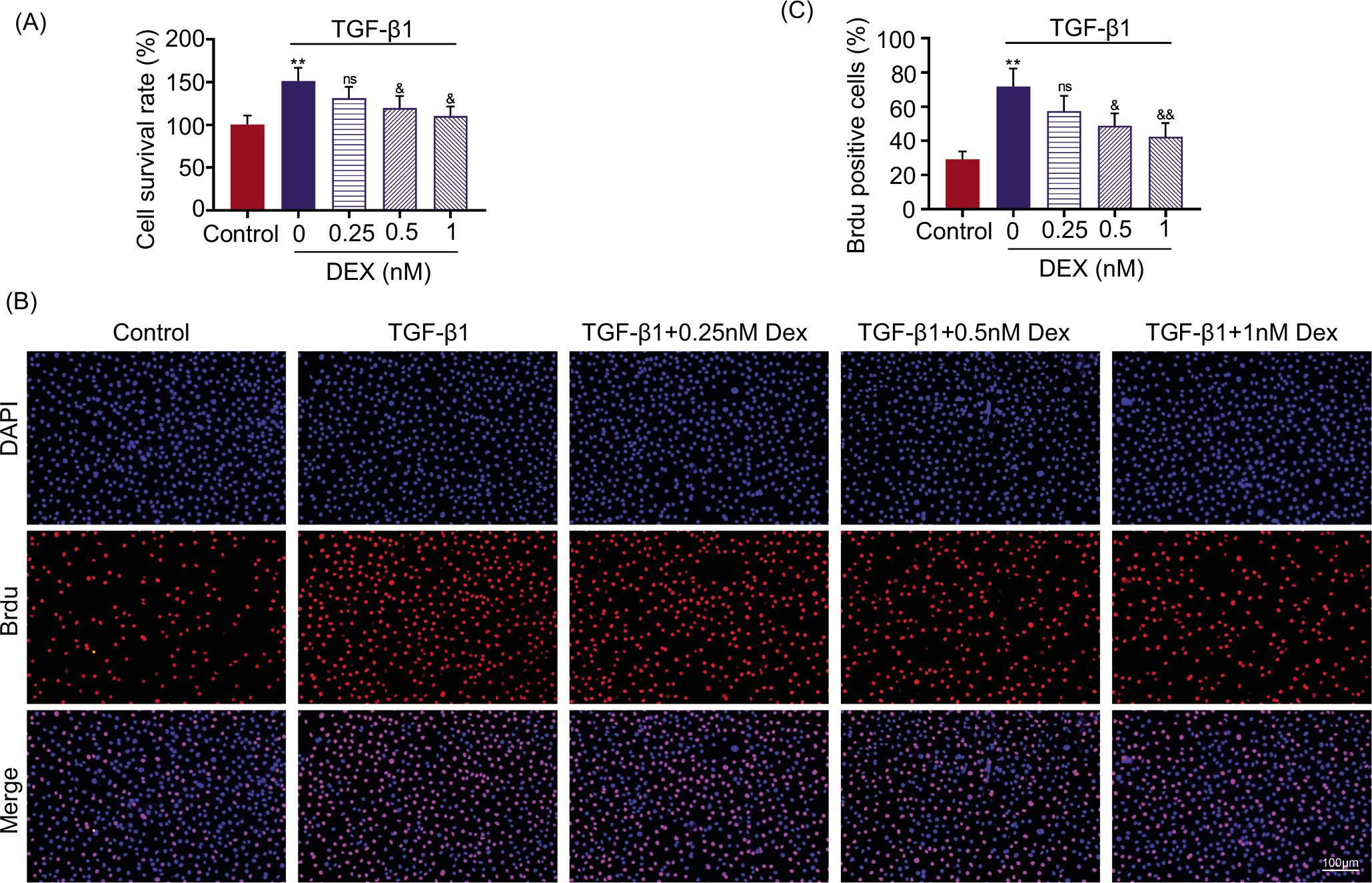
In order to establish asthmatic cell model, ASMCs were treated with TGF-β1. TGF-β1 significantly increased the cell viability of ASMCs (P < 0.01; Figure 1A), and promoted cell proliferation (P < 0.01; Figure 1B) through up-regulation of BrdU positive cells (Figure 1C). Dexmedetomidine incubation reduced cell viability of TGF-β1-induced ASMCs in a dosage-dependent manner (Figure 1A), and repressed cell proliferation (Figures 1B and 1C). Therefore, dexmedetomidine decreased proliferation of TGF-β1-induced ASMCs.
Figure 1 Dexmedetomidine suppressed cell proliferation of TGF-β1-induced ASMCs. (A) Dexmedetomidine incubation reduced cell viability of TGF-β1-induced ASMCs measured by MTT assay. (B) Dexmedetomidine incubation repressed the cell proliferation of TGF-β1-induced ASMCs observed via reduction in BrdU positive cells captured by laser scanning cytometry. (C) Dexmedetomidine reduced the number of BrdU positive cells in TGF-B1-induced ASMCs in a dose-dependent manner. N = 3. Dunnett’s test was performed for statistical analysis. **vs. control, P < 0.01. &, &&vs. 0-nM dexmedetomidine (DEX), P < 0.05, P < 0.01.
Dexmedetomidine promoted cell apoptosis of TGF-β1-induced ASMCs
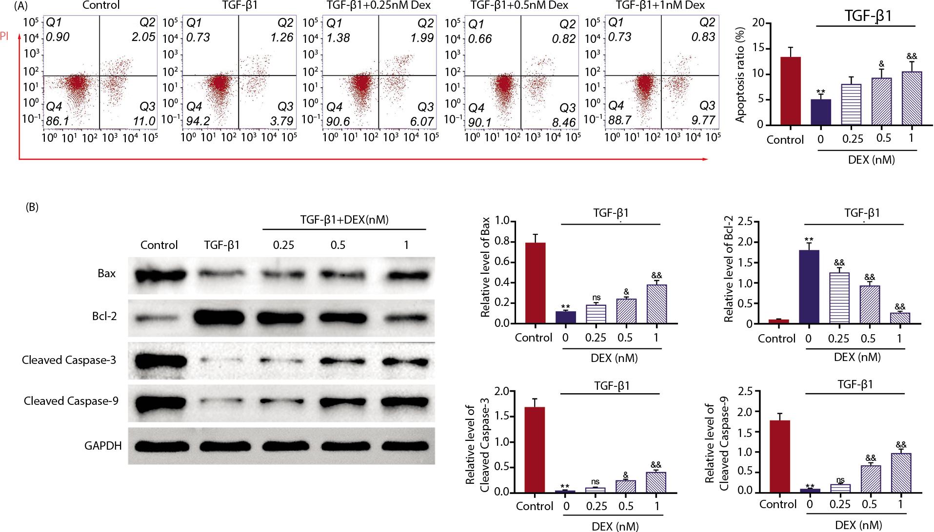
Cell apoptosis of ASMCs was significantly repressed by TGF-β1 condition (P < 0.01; Figure 2A), while dexmedetomidine promoted the apoptosis of TGF-β1-induced ASMCs in a dosage-dependent manner (Figure 2A). TGF-β1-induced increase in Bcl-2 expression and decrease in Bax-cleaved caspase-3 and cleaved caspase-9 expression in ASMCs were reversed by incubation of dexmedetomidine (Figure 2B), suggesting the pro-apoptotic role of dexmedetomidine in TGF-β1-induced ASMCs.
Figure 2 Dexmedetomidine promoted cell apoptosis of TGF-β1-induced ASMCs. (A) TGF-β1-induced ASMCs with or without dexmedetomidine treatment were incubated with 0.5 μL of Annexin-V-FITC for 10 min and 10 μL of propidium iodide (PI) (1 mg/mL) for 30 min and then analyzed by flow cytometry analysis. Results established that dexmedetomidine promoted cell apoptosis of TGF-β1-induced ASMCs. (B) Dexmedetomidine decreased Bcl-2 and increased Bax, cleaved caspase-3, and cleaved caspase-9 normalized to GAPDH in TGF-β1-induced ASMCs. N = 3. Dunnett’s test was performed for the statistical analysis. **vs. control, P < 0.01. &, &&vs. 0-nM dexmedetomidine (DEX), P < 0.05, P < 0.01.
Dexmedetomidine repressed extracellular matrix production of TGF-β1-induced ASMCs
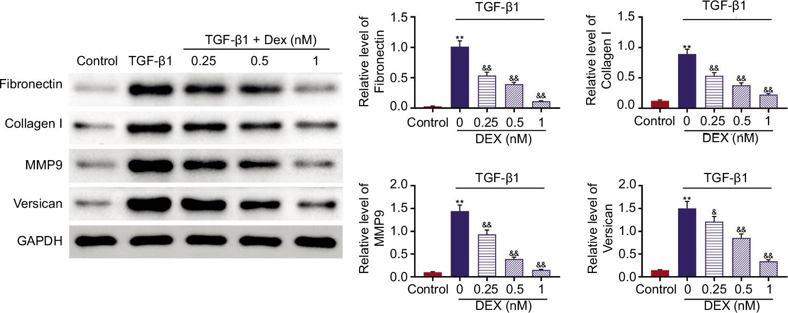
Protein expression levels of fibronectin, collagen I, MMP9, and versican were significantly up-regulated in ASMCs post-TGF-β1 condition (P < 0.01; Figure 3). However, incubation of dexmedetomidine reduced expression of fibronectin, collagen I, MMP9, and versican in TGF-β1-induced ASMCs in a dosage-dependent manner (Figure 3), demonstrating that dexmedetomidine protected ASMCs against TGF-β1-induced ECM deposition.
Figure 3 Dexmedetomidine repressed extracellular matrix (ECM) production of TGF-β1-induced ASMCs. Dexmedetomidine incubation reduced expression of fibronectin, collagen I, MMP9, and versican normalized to GAPDH in TGF-β1-induced ASMCs. N = 3. Dunnett’s test was performed for statistical analysis. **vs. control, P < 0.01. &, &&vs. 0-nM de xmedetomidine (DEX), P < 0.05, P < 0.01.
Dexmedetomidine repressed activation of mitogen-activated protein kinase (MAPK) signaling in TGF-β1-induced ASMCs
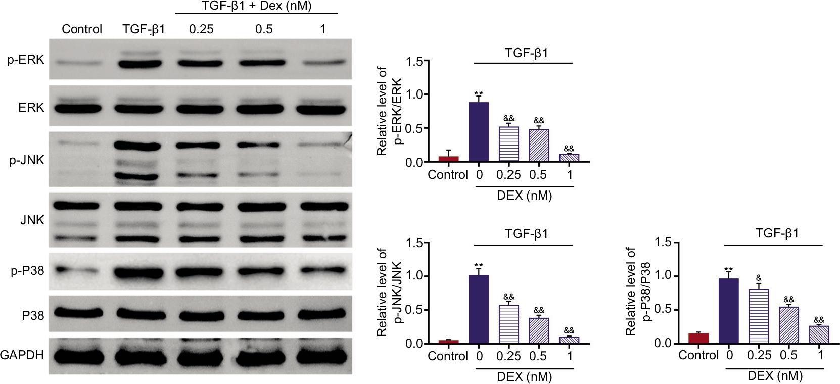
Although protein expression levels of JNK, ERK, and p38 were not affected by TGF-β1 condition (Figure 4), p-JNK, p-ERK, and p-p38 were significantly up-regulated in TGF-β1-induced ASMCs (P < 0.01; Figure 4). Moreover, incubation of dexmedetomidine decreased expression of p-JNK, p-ERK, and p-p38 in TGF-β1-induced ASMCs in a dosage-dependent manner (Figure 4), indicating the suppressive effect of dexmedetomidine on MAPK signaling in TGF-β1-induced ASMCs.
Figure 4 Dexmede tomidine repressed activation of MAPK signaling in TGF-β1-induced ASMCs. Dexmedetomidine incubation decreased expression of p-JNK, p-ERK, and p-p38 normalized to GAPDH in TGF-β1-induced ASMCs. N = 3. Dunnett’s test was performed for the statistical analysis. **vs. control, P < 0.01. &, &&vs. 0-nM dexmedetomidine (DEX), P < 0.05, P < 0.01.
Discussion
In the present study, our results demonstrated that dexmedetomidine suppressed cell proliferation of TGF-β1-induced ASMCs, promoted cell apoptosis, and repressed production of ECM. Mechanistic study indicated that dexmedetomidine repressed the activation of MAPK signaling in TGF-β1-induced ASMCs.
Previous study has established that α2-adrenoceptors bind to endogenous or exogenous agonists to mediate various endocrine, behavioral, and physiological functions, thus implicating in the pathogenesis of various diseases, such as cognitive functions, endogenous depression, hypertension, and anxiety.22 Antagonist of α2-adrenoceptor, midaglizole, was associated with bronchial hyperresponsiveness in patients with mild asthma.23 Dexmedetomidine functions as an α2-adrenoceptor agonist and repressed histamine-induced bronchoconstriction, thus benefiting in the decrease of airway reactivity in asthmatic patients.24 Dexmedetomidine was administered to treat acute asthma (as an adjunctive treatment). Moreover, increasing evidence has proved that TGF-β1, increased in asthma, modulated airway remodeling through promoting proliferation of ASMCs.25 The proliferative effect of TGF-β1 on ASMCs was first ascertained in this study as demonstrated by increased cell viability and proliferation of ASMCs after TGF-β1 treatment, and TGF-β1-repressed cell apoptosis of ASMCs. Dexmedetomidine has been reported to enhance expression of miR-21 and reduce expression of programmed cell death protein 4 to repress the development of abdominal aortic aneurysm,26 and TGF-β1 up-regulated miR-181a to decrease phosphatase and tensin homologue deleted on chromosome ten (PTEN) in asthmatic mice.27 Therefore, dexmedetomidine might regulate miR-181a/PTEN to be implicated in the TGF-β1-induced ECM production and proliferation of ASMCs.
TGF-β1 also promoted ECM production via activation of intracellular mediators, Smad proteins.28 In this study, our results demonstrated that TGF-β1 induced protein expression of fibronectin, collagen I, MMP9, and versican in ASMCs, thus promoting ECM deposition. Previous study has demonstrated the pro-apoptotic effect of dexmedetomidine on esophageal cancer cells.29 The components of ECM, including Tenascin C, matrix metallopeptidase 16, collagen IV, and fibronectin, were reduced in breast cancer cells by dexmedetomidine condition.30 Results in this study established that dexmedetomidine incubation suppressed cell proliferation of TGF-β1-induced ASMCs, promoted cell apoptosis, and reduced ECM deposition by down-regulating fibronectin, collagen I, MMP9, and versican. These results indicated that dexmedetomidine protected ASMCs against TGF-β1-induced ECM deposition and proliferation, thus alleviating airway remodeling in the development of asthma. However, TGF-β1 also contributes to airway remodeling through induction of ASMCs migration,31 and dexmedetomidine suppressed the cell migration of esophageal cancer.29 The effect of dexmedetomidine on the migration of TGF-β1-induced ASMCs must be investigated in the future research.
Mitogen-activated protein kinase signaling cascades are responsible for migration, degranulation, proliferation, activation, and differentiation of ASMCs and immune cells, and modulate airway remodeling in the development of asthma.32 MAPKs are regarded as potential targets for the treatment of asthma.33 TGF-β1 has been demonstrated to induce activation of MAPK in ASMCs, thereby promoting the proliferation, migration, and ECM deposition of ASMCs.34 Inhibition of MAPK prevented airway remodeling.35 Studies have proved that dexmedetomidine inhibited activation of MAPK signaling to protect against lidocaine-induced cytotoxicity,36 isoflurane-induced neuroapoptosis,37 and repressed ovarian cancer growth.38 Here, dexmedetomidine attenuated TGF-β1-induced increase in p-JNK, p-ERK, and p-p38 in ASMCs, thus inhibiting the activation of MAPKs.
Conclusion
The current study indicated that dexmedetomidine retarded airway remodeling in asthma through suppression of ECM deposition and proliferation in TGF-β1-induced ASMCs. Inactivation of MAPK signaling was involved in dexmedetomidine-suppressed airway remodeling. Therefore, dexmedetomidine has the potential to be used clinically for the prevention of asthma. However, there are limitations to the current study. The effects of dexmedetomidine on ASMCs migration must be studied and the ovalbum-induced asthmatic animal model must be established in the future studies to investigate in vivo effects of dexmedetomidine on asthma.
Competing Interests
The authors state that there are no conflicts of interest to disclose.
Contribution of Authors
Rong Zhou and Xiaoyan Chen designed the study and supervised data collection. Rong Zhou analyzed and interpreted the data. Xiaoyan Chen prepared the manuscript for publication and reviewed draft of the manuscript. Both authors read and approved the final manuscript.
REFERENCES
1. Mims JW, editor Asthma: definitions and pathophysiology. Int Forum Allergy Rhinol. 2015;5:S2–6.Wiley Online Library. 10.1002/alr.21609
2. Hough KP, Curtiss ML, Blain TJ, Liu R-M, Trevor J, Deshane JS, et al. Airway remodeling in asthma. Front Med (Lausanne). 2020;7:191. 10.3389/fmed.2020.00191
3. Bentley JK, Hershenson MB. Airway smooth muscle growth in asthma: Proliferation, hypertrophy, and migration. Proc Am Thorac Soc. 2008;5(1):89–96. 10.1513/pats.200705-063VS
4. Prabhala P, Wright DB, Robbe P, Bitter C, Pera T, Ten Hacken NH, et al. Laminin α4 contributes to airway remodeling and inflammation in asthma. Am J Physiol Lung Cell Mol Physiol. 2019;317(6):L768–77. 10.1152/ajplung.00222.2019
5. Dai Y, Li F, Wu L, Wang R, Li P, Yan S, et al. Roxithromycin treatment inhibits TGF-β1-induced activation of ERK and AKT and down-regulation of Caveolin-1 in rat airway smooth muscle cells. Resp Res. 2014;15:96. 10.1186/s12931-014-0096-z
6. Al-Alawi M, Hassan T, Chotirmall SH. Transforming growth factor β and severe asthma: A perfect storm. Resp Med. 2014;108(10):1409–23. 10.1016/j.rmed.2014.08.008
7. Shen Z-J, Esnault S, Rosenthal LA, Szakaly RJ, Sorkness RL, Westmark PR, et al. Pin1 regulates TGF-β1 production by activated human and murine eosinophils and contributes to allergic lung fibrosis. J Clin Invest. 2008;118(2):479–90. 10.1172/JCI32789
8. Aron J, Akbari O. Regulatory T cells and type 2 innate lymphoid cell-dependent asthma. Allergy. 2017;72(8):1148–55. 10.1111/all.13139
9. Baarsma HA, Menzen MH, Halayko AJ, Meurs H, Kerstjens HAM, Gosens R. β-Catenin signaling is required for TGF-β1-induced extracellular matrix production by airway smooth muscle cells. Am J Physiol Lung Cell Mol Physiol. 2011;301(6):L956–65. 10.1152/ajplung.00123.2011
10. Chen G, Khalil N. TGF-β1 increases proliferation of airway smooth muscle cells by phosphorylation of map kinases. Resp Res. 2006;7(1):2. 10.1186/1465-9921-7-2
11. Yang Z, Qu Z, Yi M, Lv Z, Wang Y, Shan Y, et al. MiR-204-5p inhibits transforming growth factor-β1-induced proliferation and extracellular matrix production of airway smooth muscle cells by regulating Six1 in Asthma. Int Arch Allergy Immunol. 2020;181(4):239–48. 10.1159/000505064
12. Lee S. Dexmedetomidine: Present and future directions. Korean J Anesthesiol. 2019;72(4):323–30. 10.4097/kja.19259
13. Si Y, Bao H, Han L, Chen L, Zeng L, Jing L, et al. Dexmedetomidine attenuation of renal ischaemia-reperfusion injury requires sirtuin 3 activation. Br J Anaesth. 2018; 121(6):1260-1271.10.1016/j.bja.2018.07.007
14. Xu Z, Wang D, Zhou Z, Chen Q, Zhang D, Chen S, et al. Dexmedetomidine attenuates renal and myocardial ischemia/reperfusion injury in a dose-dependent manner by inhibiting inflammatory response. Ann Clin Lab Sci. 2019;49:31–5.
15. Yuan M, Meng X-W, Ma J, Liu H, Song S-Y, Chen Q-C, et al. Dexmedetomidine protects H9c2 cardiomyocytes against oxygen-glucose deprivation/reoxygenation-induced intracellular calcium overload and apoptosis through regulating FKBP12. 6/RyR2 signaling.Drug Des Devel Ther. 2019;13:3137–49. 10.2147/DDDT.S219533
16. Zhu C, Zhou Q, Luo C, Chen Y. Dexmedetomidine protects against oxygen-glucose deprivation-induced injury through inducing astrocytes autophagy via TSC2/mTOR pathway. Neuromol Med. 2020;22(2):210–7. 10.1007/s12017-019-08576-0
17. Chen Y, Li L, Zhang J, Cui H, Wang J, Wang C, et al. Dexmedetomidine alleviates lipopolysaccharide-induced hippocampal neuronal apoptosis via inhibiting the p38 MAPK/c-Myc/CLIC4 signaling pathway in rats. Mol Neurobiol. 2021. 58(11):5533-5547. 10.1007/s12035-021-02512-9
18. Hong J, Chen Q, Wang Y, Lin S, Su Y. Dexmedetomidine alleviates smoke-induced bronchial and alveolar epithelial cell injury. Gen Physiol Biophy. 2020;39 3:293–300. 10.4149/gpb_2020003
19. Cozzi G, Lega S, Giorgi R, Barbi E. Intranasal dexmedetomidine sedation as adjuvant therapy in acute asthma exacerbation with marked anxiety and agitation. Ann Emerg Med. 2017;69(1):125–7. 10.1016/j.annemergmed.2016.08.005
20. Takasaki Y, Kido T, Semba K. Dexmedetomidine facilitates induction of noninvasive positive pressure ventilation for acute respiratory failure in patients with severe asthma. J Anesth. 2009;23:314. 10.1007/s00540-008-0712-5;10.1007/s00540-009-0762-3
21. Dai Y, Li F, Wu L, Wang R, Li P, Yan S, et al. Roxithromycin treatment inhibits TGF-β1-induced activation of ERK and AKT and down-regulation of Caveolin-1 in rat airway smooth muscle cells. Respir Res. 2014;15(1):1–8. 10.1186/s12931-014-0096-z
22. Ma D, Rajakumaraswamy N, Maze M. α2-Adrenoceptor agonists: Shedding light on neuroprotection? Br Med Bull. 2005;71(1):77–92. 10.1093/bmb/ldh036
23. Sakai H, Dobashi K, Nakazawa T. Effect of an α2-adrenoceptor antagonist, midaglizole, on bronchial responsiveness to histamine in patients with mild asthma. J Asthma. 1995;32(4):259–64. 10.3109/02770909509044833
24. Groeben H, Mitzner W, Brown Robert H. Effects of the α2-adrenoceptor agonist dexmedetomidine on bronchoconstriction in dogs. Anesthesiology. 2004;100(2):359–63. 10.1097/00000542-200402000-00026
25. Zhang H, Yan HL, Li XY, Guo YN. TNFSF14, a novel target of miR-326, facilitates airway remodeling in airway smooth muscle cells via inducing extracellular matrix protein deposition and proliferation. Kaohsiung J Med Sciences. 2020;36(7):508–14. 10.1002/kjm2.12197
26. Yu Q, Li Q, Yang X, Liu Q, Deng J, Zhao Y, et al. Dexmedetomidine suppresses the development of abdominal aortic aneurysm by downregulating the mircoRNA-21/PDCD 4 axis. Int J Mol Med. 2021;47(5):1–11. 10.3892/ijmm.2021.4923
27. Lv X, Li Y, Gong Q, Jiang Z. TGF-β1 induces airway smooth muscle cell proliferation and remodeling in asthmatic mice by up-regulating miR-181a and suppressing PTEN. Int J Clin Exp Pathol. 2019;12(1):173.
28. Laping N, Grygielko E, Mathur A, Butter S, Bomberger J, Tweed C, et al. Inhibition of transforming growth factor (TGF)-β1-induced extracellular matrix with a novel inhibitor of the TGF-β type I receptor kinase activity: SB-431542. Mol Pharmacol. 2002;62(1):58–64. 10.1124/mol.62.1.58
29. Zhang P, He H, Bai Y, Liu W, Huang L. Dexmedetomidine suppresses the progression of esophageal cancer via miR-143-3p/epidermal growth factor receptor pathway substrate 8 axis. Anticancer Drugs. 2020;31(7):693–701. 10.1097/CAD.0000000000000934
30. Chi M, Shi X, Huo X, Wu X, Zhang P, Wang G. Dexmedetomidine promotes breast cancer cell migration through Rab11-mediated secretion of exosomal TMPRSS2. Ann Transl Med. 2020;8(8):531. 10.21037/atm.2020.04.28
31. Chen M, Shi J-T, Lv Z-Q, Huang L-J, Lin X-L, Zhang W, et al. Triptolide inhibits transforming growth factor-β1-induced proliferation and migration of rat airway smooth muscle cells by suppressing nuclear factor-κB but not extracellular signal-regulated kinase 1/2. Immunology. 2015;144(3):486–94. 10.1111/imm.12396
32. Manzoor MK, Eric BP. The role of mitogen-activated protein kinases in asthma. Curr Immunol Rev. 2015;11(2):132–46. 10.2174/1573395511666150615225641
33. Khorasanizadeh M, Eskian M, Gelfand E, Rezaei N. Mitogen-activated protein kinases as therapeutic targets for asthma. Pharmacol Ther. 2017; 174:112-126. 10.1016/j.pharmthera.2017.02.024
34. Dai Y, Li F, Wu L, Wang R, Li P, Yan S, et al. Roxithromycin treatment inhibits TGF-β1-induced activation of ERK and AKT and down-regulation of Caveolin-1 in rat airway smooth muscle cells. Resp Res. 2014;15(1):96. 10.1186/s12931-014-0096-z
35. Sun Y, Shi Z, Liu B, Li XG, Li G, Yang F, et al. YKL-40 mediates airway remodeling in asthma via activating FAK and MAPK signaling pathway. Cell Cycle. 2020;19(11):1378–90. 10.1080/15384101.2020.1750811
36. Wang Q, Tan Y, Zhang N, Xu Y, Wei W, She Y, et al. Dexmedetomidine inhibits activation of the MAPK pathway and protects PC12 and NG108-15 cells from lidocaine-induced cytotoxicity at its maximum safe dose. Biomed Pharmacother. 2017;91:162–6. 10.1016/j.biopha.2017.04.084
37. Liao Z, Cao D, Han X, Liu C, Peng J, Zuo Z, et al. Both JNK and P38 MAPK pathways participate in the protection by dexmedetomidine against isoflurane-induced neuroapoptosis in the hippocampus of neonatal rats. Brain Res Bull. 2014;107:69–78. 10.1016/j.brainresbull.2014.07.001
38. Cai Q-H, Tang Y, Fan S-H, Zhang Z-F, Li H, Huang S-Q, et al. In vivo effects of dexmedetomidine on immune function and tumor growth in rats with ovarian cancer through inhibiting the p38MAPK/NF-κB signaling pathway. Biomed Pharmacother. 2017;95:1830–7. 10.1016/j.biopha.2017.09.086