
Download
ORIGINAL ARTICLE
Fabrication and production of conjugated neurotensin–silver nanoparticles and evaluation of its effect on pathophysiology of allergic asthma
Yu Yanga, Entezar Mehrabi Nasabb, Seyyed Shamsadin Atharic*
aDepartment of Respiratory, The Second Affiliated Hospital Zhejiang University School of Medicine, Hangzhou, 310009, China
bDepartment of Cardiology, School of Medicine, Mousavi Hospital, Zanjan University of Medical Sciences, Zanjan, Iran
cDepartment of Immunology, School of Medicine, Zanjan University of Medical Sciences, Zanjan, Iran
Abstract
Asthma, a respiratory tract disease, is characterized by inflammation and obstruction of airway. Inflammatory cells play a significant role in allergic asthma, and there is no complete cure for asthma. One of the new approaches in medicines is nanoparticle-base treatment. The aim of the current study is to introduce a new therapeutic approach in nano-medicine with neurotensin. Conjugated peptide nanoparticles were prepared and characterized, and then administrated to asthmatic mice. Airway hyperresponsiveness (AHR) test, broncho-alveolar lavage fluid (BALF) cells counting, cytokines level, and histopathology study were conducted. Treatment with peptide nanoparticles could control AHR, percentage of eosinophils in BALF, levels of interleukin 4 (IL-4), IL-5, and IL-33, peri-airways and perivascular eosinophilic inflammation. Producing and using of new peptide nano-drugs could introduce new therapeutic approach in controlling pathological-related mechanisms in allergic asthma.
Key words: allergy, inflammation, lung, nanoparticle
*Corresponding author: S.S. Athari, Department of Immunology, School of Medicine, Zanjan University of Medical Sciences, Zanjan, Iran. Email address: [email protected]; [email protected]
Received 21 April 2025; Accepted 11 June 2025; Available online 1 September 2025
Copyright: Yang Y, et al.
This open access article is licensed under Creative Commons Attribution 4.0 International (CC BY 4.0). http://creativecommons.org/licenses/by/4.0/
Introduction
Asthma, one of the non-communicable diseases of respiratory tract, is characterized by inflammation and obstruction of the airway. More than 300 hundred million people are suffering from asthma globally, and it has become a huge economic burden of state as well as families.1–4 Asthma symptoms, such as difficult and short breathing, wheezing, high heart rate, and chest pain, result from inflammation in the airway, which triggers processes such as increased production of mucus, changes in the structure of airway walls, and bronchial hyperresponsiveness (BHR). Asthma is caused by a combination of intricate and unknown genetic and environmental factors, and these factors are important in both severity of the disease and response to its treatment. Family history and various genes are involved as asthma risk factors; these genes are associated with immune system and inflammatory responses. Inflammatory cells play a significant role in allergic asthma by secreting pro-inflammatory and inflammatory cytokines, such as interleukin-4 (IL-4), IL-5, IL-9, IL-13, IL-17, IL-25, and IL-33. Although there is no cure for asthma, its manifestations could be controlled. The most effective treatment for asthma is to detect its triggers and eliminate exposure to the same combined with medicines to control bronchial inflammation.5–9
One of the new approaches in discovering and creating therapies is nanoparticle-based treatment. Nano-drugs are improvement of therapeutic agents using nanotechnology. Recently, nano-medicine has received a lot of attention and is used to improve drug delivery in the body. Metal nanoparticles are used widely, but gold and silver are the most widely used elements, and different ligands, such as peptides, can bind to the particles.10–12
Neurotensin, a 13-amino acid peptide, as a hormone and neurotransmitter, was first isolated from bovine hypothalamus that influences gut motility. Neurotensin is an important regulator of immune cells and acts as a linker between the immune and nervous systems. This neuroimmune interaction is observed in asthma and neurotensin affects as a modulator in both inflammatory cells and lung nerves.13 The main aim of the current study is to introduce a new therapeutic approach in nano-medicine to study and control pathophysiology of allergic asthma, and enhance immunomodulatory activity with endogenous peptide through nanoparticles.
Materials and Methods
Peptide nanoparticle preparation
Linker molecules were used as a covalent bonding to bind neurotensin to silver nanoparticles. Briefly, aldehyde active groups of neurotensin and hydrazine active groups of silver nanoparticles were conjugated by connecting these two active groups. Then, a high-speed centrifuge was used to ensure the complete purification of peptide-bound nanoparticles.11,12 Scanning electron microscopy (SEM) and Fourier Transform Infrared Spectroscopy (FTIR) spectroscopy were applied to confirm the formation of conjugated peptide nanoparticles.
Peptide releasing was investigated at pH 7.4 (physiological), 7, and 6.4 (similar to bronchi pH), and concentration of the released peptide was determined according to standard curve.11,12
Allergic asthma model
Allergic asthma model for the study was prepared using BALB/c mice by administering ovalbumin (OVA) with alum adjuvant through intraperitoneal route (on days 1 and 14) and repeating through inhalation for sensitization and challenging by 1% of OVA solution (on days 24, 26, 28, and 30) as described in the literature.3,8,12,14 Under standard animal care conditions, mice were allocated in the following five groups: asthma group (group A), healthy control group, which received phosphate-buffered saline (PBS) only (group B), and three allergic asthma groups that received silver nanoparticle (group C), neurotensin peptide (group D), and neurotensin–silver nanoparticle (group E).. On day 30, airway hyperresponsiveness (AHR) test was conducted and on day 31, the mice were euthanized by CO2 and then sampled.
AHR test
Airway hyperresponsiveness was performed in all groups according to previously described method.3,8,12,14,15 In brief, metacholine (Mch) challenge test was done in increased dose of Mch (0, 1, 2, 4, 8, 16, and 32 mg/mL) via nebulizing and AHR was assessed as Penh (or enhanced pause) value.
BAL fluid cells
Broncho-alveolar lavage fluid (BALF) was collected and cytospine slides were prepared to determine eosinophil percentage. The supernatant of BALF was used to measurement of bio-factors.
Luminex assay
A multiplex mouse cytokine, chemokine, and growth factor detection kit were used to measure levels of cytokines, such as IL-4, IL-5, IL-13, and IL-33, according to manufacturer’s description.
Histopathology
Lung tissues of mice were isolated and fixed. The histopathology sections were stained with hematoxyline and eosin (H&E), Periodic acishiff (PAS), and Alcian blue (AB)–PAS. The slides were evaluated under microscopy to determine mucus produced in the airways, goblet cell hyper/metaplasia (an abnormal increase in the number of goblet cells), and eosinophilic inflammation in perivascular (around blood vessels) and peri-airways (around airways) of the lungs.3,8,12,14,16
Statistical analysis
The result was expressed as means ± SD. Differences between studied groups were analyzed with a t-test, and P < 0.05 was considered statistically significant. GraphPad Prism was used to present the data.
Result
Nanoparticles
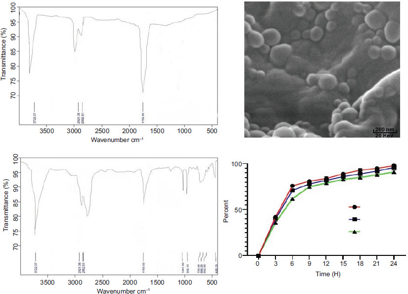
The SEM figure described semi-spheral shape of nanoparticles that had an average size of 142 ± 6 nm. FTIR spectroscopy presented and confirmed the conjugation of peptide nanoparticles. Assays performed at pH 7.4, 7, and 6.4 showed that a large amount of peptide releasing occurred in first 6 h (Figure 1).
Figure 1 SEM of neurotensin conjugated to silver nanoparticles has semi-spheral shape. FTIR confirmed the formation of the conjugated peptide-nanoparticle. Also, peptide releasing percentage was measured in several pH; 7.4, 7, and 6.4 (in 24 h).
AHR
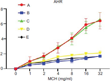
AHR results showed that Penh value in asthma group increased significantly (P < 0.05), compared to healthy group in response to Mch concentration (Figure 2). In addition, group C was similar to asthma group (group A), but in the other three groups, AHR decreased significantly (P < 0.05), compared to the asthma group.
Figure 2 Study of airway hyperresponsiveness in all groups was done through Mch challenge test.
BALF eosinophil
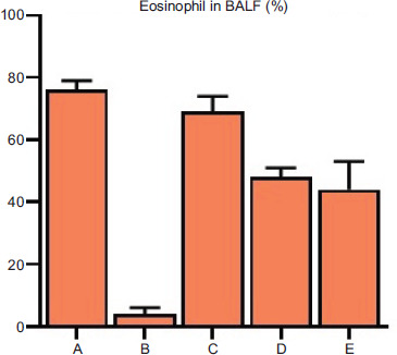
The percentage of eosinophils was elevated in the BALF of asthmatic mice, compared to group B (Figure 3). In addition, group C was similar to group A, but in the other two groups (groups D and E), percentage of eosinophils decreased significantly (P < 0.05), compared to group A.
Figure 3 Percentage of eosinophils was determined in broncho-alveolar lavage fluid.
Levels of cytokines
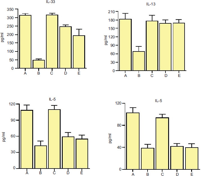
The levels of main cytokines, IL-4, IL-5, IL-33, and IL-13, were enhanced significantly (P < 0.05) in group A, compared to group B (Figure 4). Levels of these four cytokines in group C were similar to that of group A, but in the other two groups (groups D and E), levels of IL-4, IL-5, and IL-33 decreased significantly (P < 0.05), compared to group A, but decrease in the levels of IL-13 in groups D and E was not significant (P > 0.05).
Figure 4 Levels of the main allergic cytokines, IL-4, IL-5, IL-13, and IL-33, were measured in the broncho-alveolar lavage fluid of mice.
Histopathology
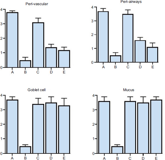
In group A, goblet cell metaplasia, excessive mucus production in the airways and peri-airways, and perivascular eosinophilic inflammation were increased significantly (P < 0.05), compared to group B (Figures 5 and 6). Goblet cell metaplasia and excessive mucus production in the airways showed no significant changes (P > 0.05) in groups C, D, and E, compared to group A. Peri-airways and perivascular eosinophilic inflammation decreased significantly (P < 0.05) in groups D and E, compared to group A, but this decrease was not significant (P > 0.05) in group C, compared to group A.
Figure 5 Mucus production, metaplasia of the goblet cell, and eosinophilic inflammation in perivascular and peribronchial were studied in pathological sections.
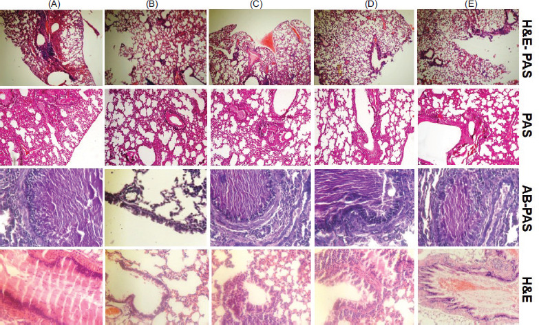
Figure 6 Lung sections of mice were prepared and stained with hematoxylin and eosin–periodic acid-Schiff (H&E-PAS), PAS, Alcian Blue–PAS (AB-PAS), and H&E.
Discussion
Asthma is a highly complicated chronic pulmonary inflammatory disease, involving a variety of cells and molecules. Therefore, it has many potential targets, including transcription factors, chemokines, tyrosine kinases, cytokines and their receptors as well as co-stimulatory molecules, which can be manipulated through nanoparticles. Traditional anti-asthmatics are administered by inhalation, intravenously, or orally. Inhaled agents are the mainstream medications to control asthma, and compared to systemically administrated drugs, the targeted drug delivery by inhalation improves the bioavailability of drugs. The drugs directly act on the respiratory tract with less dosage and fewer systemic adverse reactions and toxicity.10,17,18 The most commonly used drugs include inhaled β2-receptor agonists, anticholinergic drugs, corticosteroids, and short-acting theophylline.
The current nano-drugs are divided into two categories: nanotechnology-improved traditional molecular drugs, and brand-new nano-drugs. The nano form of traditional drugs mainly includes nanoparticle carriers developed with precise surface patterns, and the existing drugs carrying through drug-targeting reagents. Nontoxic nanoparticle, telomere dendrimer, a dendrimer molecule designed or modified to interact with or mimic telomeres), is an efficient nano-carrier with better stability and greater loading capacity that delivers hydrophobic drugs directly into the lungs, reduces allergic pulmonary inflammation, eosinophils, and inflammatory cytokines.19–21 Many vectors based on liposomes and polymers have been developed to convert nucleic acids into nanoparticles for lung delivery. Applications of nanoparticles, such as polyethyleneimine (PEI), chitosan, polyamidoamine dendrimers, and biodegradable poly(lactic-co-glycolic) acid (PLGA) in the delivery of nucleic acids to the lungs have been described.10,22–26 Therefore, selecting and producing a suitable nanoparticle as a drug carrier is important and helps to achieve good results to control asthma with the best vision and without or least adverse effects. AHR results showed that Penh value decreased due to treatment with peptides and peptide nanoparticles.
Furthermore, in allergen-specific immunotherapy, nontoxic, sterile, and endotoxin-free formulations are required for safe clinical application of nanoparticles. Sterilization of nanoparticles after synthesis may be problematic as it may alter properties.27 It was observed that proteolytic processing enhanced uptake by macropinocytosis as well as antigen presentation of allergen together with the ability to boost IgG2a antibody (a specific subclass of immunoglobulin G [Ig-G]) and diminish immunoglobulin E (IgE) levels upon SiO2 nanoparticle interactions. All events imply the skewing of immune responses toward a Th1-dominated immune profile and decreased allergic sensitization. Therefore, SiO2 nanoparticles may benefit as an efficient allergen-immunotherapy nano-carrier platform associated with a nonparticulate adjuvant.28
In a study, to overcome metabolization and clearance of the drug, targeted drug delivery based on nanotechnology was developed to increase the bioavailability of herbal drugs to treat asthma. In a mouse model of house dust mite (HDM)-induced asthma, a nano-herbal drug inhibited inflammation in the lungs as evidenced by reduced inflammatory cells and inflammatory cytokines. The prescribed nano-herbal drug markedly inhibited IL-4, IL-5, and IL-13 (Th2 cytokines, produced by Th2 cells) and elevated IL-12 levels (Th1 cytokines). IL-12 is involved in the antagonism of Th2-responses and IgE synthesis to restrain the progress of asthma.29 Levels of IL-4, IL-5, and IL-33 decreased significantly in two treated groups (groups D and E), compared to non-treated asthma group.
In another study, intra-tracheal administration of mucus-penetrating nanoparticles carrying thymulin-expressing plasmids normalized the pathologic features of asthmatic lungs (including pulmonary fibrosis, chronic inflammation, and mechanical perturbation). It reduced eosinophil counts, IL-4, IL-13, and vascular endothelial growth factor (VEGF), concomitantly normalizing chemokine CCL11 (eotaxin-1) level, and blocking eosinophil recruitment and collagen deposition. It was reported that pro-fibrotic mediators and neutrophil-recruiting CXCL1 were reduced by treatment with a single dose of thymulin-expressing nanoparticles; shifted phenotypic from pathological TH2 subtype to therapeutic Treg; and the Treg-recruiting CCL17 level was elevated. Thymulin phenotypic nanoparticle deviating macrophages from M2 phenotype may be attributed to a reduction in M2-inducing TH2 cytokine production.30–32 The elevated percentage of eosinophils in the BALF of asthmatic mice was reduced in two treated groups (groups D and E).
Although some studies indicated the pro-inflammatory activity of neurotensin in the murine model of sepsis and the animal model of colitis,33,34 neurotensin signaling was up-regulated in experimental colitis during the healing process. In a study, neurotensin exhibited anti-inflammatory activity through the reduced levels of IL-6 and tumor necrosis factor-α (TNF-α) in serum and malondialdehyde, caspase-3, and myeloperoxidase in colonic tissue.13,35 Neurotensin and its receptors have been identified in airway mucosa, in presynaptic cholinergic terminals, and in post-synaptic smooth muscles of the bronchi.13,36 Neurotensin administration could reduce airway responsiveness to methacholine provocation.13 About cytokines, IL-17A has an important role in lung inflammation and is secreted by distinctive T cells of Th17 subtype. IL-17A is expressed in the BALF of asthmatic patients. It induces neutrophil chemotactic factor (CXCL8) released from airway smooth muscles and epithelial cells that leads to neutrophil recruitment. Another main cytokine in asthma is IL-13 that was reduced by neurotensin treatment.37–40 Eosinophilic inflammation in peri-airways and perivascular was significantly controlled by treatment in groups D and E, compared to group A; however, goblet cell metaplasia and excessive mucus production in the airways showed no significant changes in groups D and E, compared to group A.
Neurotensin acts as pulmonary neutral endopeptidase that leads to bronchodilation.41 Neurotensin is a modulator of different processes, including mast cells-mediated inflammation and catecholamine production, but it is quickly metabolized in pulmonary parenchyma. Neutral endopeptidase is rapidly eliminated from the circulation by metabolism.42,43 Therefore, in this study, to prevent rapid elimination of neurotensin, it was conjugated with silver nanoparticles to carry peptides into the airway. These peptides affect target cells before metabolization and have strong and better effects to control asthma symptoms.
In this research, the potential benefits of peptide nano-carriers to treat asthma were studied, and a potentially useful technique for controlling asthma symptoms and treatment of lung diseases with nanotechnology was applied. The effectiveness of peptide nano-carriers in the animal model of asthma was observed. However, many challenges still need to be overcome for applying peptide nano-medicine therapy and for understanding the mechanism of asthma pathogenesis as well as its relation with nano-carriers, important for the implementation and design of reasonable nano-based peptide therapy.
The novel drug nano-delivery system provides a promising platform for improving asthma treatment. This study has several limitations, such as the chronic form of asthma was not studied; toxicity and adverse effects of the produced nanoparticles were not evaluated; and effects of the produced component on other organs were not studied.
Ethical Approval and Consent to Participate
The current study was approved by the Ethics Committee of Animal House of ix.med.vet.dep, 2024 (No. IX.MED.VET.DEP.REC.2024.0100019.8).
Availability of Data and Materials
Data are available on request from corresponding author.
Author Contributions
All the authors participated in the designing, animal study, laboratory analysis, and writing of the manuscript.
Conflicts of Interest
There is no conflict of interest.
Funding
Not Applicable.
REFERENCES
1 Qian L, Mehrabi Nasab E, Athari, SM, Athari SS. Mitochondria signaling pathways in allergic asthma. J Investig Med. 2022;70(4):863–82. 10.1136/jim-2021-002098
2 Esmaeilzadeh A, Tahmasebi S, Athari SS. Chimeric antigen receptor-T cell therapy: Applications and challenges in treatment of allergy and asthma. Biomed Pharmacother. 2020;123:109685. 10.1016/j.biopha.2019.109685
3 Athari SM, Mehrabi Nasab E, Athari SS. Study effect of ocimumbasilicum seeds on mucus production and cytokine gene expression in allergic asthma mice model. Rev Fr Allergol. 2018;58(7):489–93. 10.1016/j.reval.2018.08.003
4 Athari SS, Athari SM. The importance of eosinophil, platelet and dendritic cell in asthma. Asian Pac J Trop Dis. 2014;4(1): 41–7. 10.1016/S2222-1808(14)60413-8
5 Lankarani KB, Honarvar B, Athari SS. The mechanisms underlying helicobacter pylori-mediated protection against allergic asthma. Tanaffos. 2017;16(4):251–9.
6 Jiang J, Nasab EM, Athari SM, Athari SS. Effects of vitamin E and selenium on allergic rhinitis and asthma pathophysiology. Resp Physiol Neurobiol. 2021;286:103614. 10.1016/j.resp.2020.103614
7 Haddadzadeh H., Athari SS, Hajimohammadi B. The first record of linguatulaserrata infection of two-humped camel (Camelusbacterinus) in Iran. Iran J Parasitol. 2009;4(1):59–61.
8 Nasab EM, Athari SM, Motlagh B, Athari SS. Effects of oral administration of ocimumbasilicum on goblet cell hyperplasia and upstream cytokine gene expression in allergic asthma. Rev Fr Allergol. 2020;60:64–68. 10.1016/j.reval.2019.02.226
9 Huang M, Nasab EM, Athari SS. Immunoregulatory effect of mesenchymal stem cell via mitochondria signaling pathways in allergic asthma. Saudi JBiol Sci. 2021;28:6957–6962. 10.1016/j.sjbs.2021.07.071
10 Wang L, Feng M, Li Q, Qiu C, Chen R. Advances in nanotechnology and asthma. Ann Transl Med. 2019;7(8):180. 10.21037/atm.2019.04.62
11 Nasab DN, Taheri A, Athari SS. Evaluation anti-inflammatory effect of conjugated gold nanoparticles with cortistatin peptide as drug delivery to asthmatic lung tissue. Int J Peptide Res Therap. 2023;29:16. 10.1007/s10989-022-10487-x
12 Athari SS, Pourpak Z, Folkerts G, Garssen J, Moin M, Adcock IM, et al. Conjugated alpha-alumina nanoparticle with vasoactive intestinal peptide as a nano-drug in treatment of allergic asthma in mice. Eur J Pharmacol. 2016;791:811–20. 10.1016/j.ejphar.2016.10.014
13 Russjan E, Kaczynska K. Beneficial effects of neurotensin in murine model of hapten-induced asthma. Int J Mol Sci. 2019;20:5025. 10.3390/ijms20205025
14 Wang D, Nasab EM, Athari SS. Study effect of baicalein encapsulated/loaded chitosan-nanoparticle on allergic asthma pathology in mouse model. Saudi J Biol Sci. 2021;28:4311–7. 10.1016/j.sjbs.2021.04.009
15 Arora P, Athari SS, Nainwal LM. Piperine attenuates production of inflammatory biomarkers, oxidative stress and neutrophils in lungs of cigarette smoke-exposed experimental mice. Food Biosci. 2022;49:101909. 10.1016/j.fbio.2022.101909
16 Nasaba EM, Atharib SM, Ghafarzadec S, Nasabd ARM, Athari SS. Immunomodulatory effects of two silymarin isomers in a Balb/c mouse model of allergic asthma. Allergol Immunopathol. 2020;48(6):646–53. 10.1016/j.aller.2020.01.003
17 Nasr M, Najlah M, D’Emanuele A, Elhissi A. PAMAM dendrimers as aerosol drug nanocarriers for pulmonary delivery via nebulization. Int J Pharm. 2014;461:242–50. 10.1016/j.ijpharm.2013.11.023
18 Nasab EM, Makoei RHZ, Aghajani H, Athari SS. IL-33/ST2 pathway as upper-hand of inflammation in allergic asthma contributes as predictive biomarker in heart failure. ESC Heart Fail. 2022;9(6):3785–90. 10.1002/ehf2.14111
19 Kenyon NJ, Bratt JM, Lee J, Luo J, Franzi LM, Zeki AA, et al. Self-assembling nanoparticles containing dexamethasone as a novel therapy in allergic airways inflammation. PLoS One. 2013;8:e77730. 10.1371/journal.pone.0077730
20 Jackson JK, Zhang X, Llewellen S, Hunter WL, Burt HM. The characterization of novel polymeric paste formulations for intratumoral delivery. Int J Pharm. 2004;270:185–98. 10.1016/j.ijpharm.2003.10.010
21 Bao X-H, Gao F, Athari SS, Wang H. Immunomodulatory effect of IL-35 gene-transfected mesenchymal stem cells on allergic asthma. Fundam Clin Pharmacol. 2023;37(1):116–24. 10.1111/fcp.12823
22 Di Gioia S, Trapani A, Castellani S, Carbone A, Belgiovine G, Craparo EF, et al. Nanocomplexes for gene therapy of respiratory diseases: Targeting and overcoming the mucus barrier. Pulm Pharmacol Ther. 2015;34:8–24. 10.1016/j.pupt.2015.07.003
23 Merdan T, Callahan J, Petersen H, Kunath K, Bakowsky U, Kopecková P, et al. Pegylatedpolyethylenimine-Fab′ antibody fragment conjugates for targeted gene delivery to human ovarian carcinoma cells. Bioconjug Chem. 2003;14:989–96. 10.1021/bc0340767
24 Köping-Höggård M, Tubulekas I, Guan H, Edwards K, Nilsson M, Vårum KM, et al. Chitosan as a nonviral gene delivery system. Structure-property relationships and characteristics compared with polyethylenimine in vitro and after lung administration in vivo. Gene Ther. 2001;8:1108–21. 10.1038/sj.gt.3301492
25 Rudolph C, Lausier J, Naundorf S, Müller RH, Rosenecker J. In vivo gene delivery to the lung using polyethylenimine and fractured polyamidoamine dendrimers. J Gene Med. 2000;2:269–78. 10.1002/1521-2254(200007/08)2:4<269::AID-JGM112>3.0.CO;2-F
26 Bivas-Benita M, Lin MY, Bal SM, Meijgaarden KEV, Franken KLMC, Friggen AH, et al. Pulmonary delivery of DNA encoding mycobacterium tuberculosis latency antigen Rv1733c associated to PLGA-PEI nanoparticles enhances T cell responses in a DNA prime/protein boost vaccination regimen in mice. Vaccine. 2009;27:4010–7. 10.1016/j.vaccine.2009.04.033
27 Pohlit H, Bellinghausen I, Frey H, Saloga J. Recent advances in the use of nanoparticles for allergen-specific immunotherapy. Allergy. 2017;72:1461–74. 10.1111/all.13199
28 Johnson L, Aglas L, Punz B, Dang H-H, Christ C, Pointner L, et al. Mechanistic insights into silica nanoparticle–allergen interactions on antigen presenting cell function in the context of allergic reactions. Nanoscale. 2023;15:2262. 10.1039/D2NR05181H
29 Jin H, Li J, Zhang M, Luo R, Lu P, Zhang W, et al. Berberine-loaded biomimetic nanoparticles attenuate inflammation of experimental allergic asthma via enhancing IL-12 Expression. Front Pharmacol. 2021;12:724525. 10.3389/fphar.2021.724525
30 da Silva AL, de Oliveira GP, Kim N, Cruz FF, Kitoko JZ, Blanco NG, et al. Nanoparticle-based thymulin gene therapy therapeutically reverses key pathology of experimental allergic asthma. Sci Adv. 2020;6:eaay7973. 10.1126/sciadv.aay7973
31 Lunin SM, Khrenov MO, Novoselova TV, Parfenyuk SB, Novoselova EG. Thymulin, a thymic peptide, prevents the overproduction of pro-inflammatory cytokines and heat shock protein Hsp70 in inflammation-bearing mice. Immunol Invest. 2008;37:858–70. 10.1080/08820130802447629
32 Lunin SM, Glushkova OV, Khrenov MO, Parfenyuk SB, Novoselova TV, Fesenko EE, et al. Thymus peptides regulate activity of RAW 264.7 macrophage cells: Inhibitory analysis and a role of signal cascades. Expert Opin Ther Targets. 2011;15:1337–46. 10.1517/14728222.2011.641952
33 Piliponsky AM, Chen CC, Nishimura T, Metz M, Rios EJ, Dobner PR, et al. Neurotensin increases mortality and mast cells reduce neurotensin levels in a mouse model of sepsis. Nat Med. 2008;14:392–8. 10.1038/nm1738
34 Law IK, Bakirtzi K, Polytarchou C, Oikonomopoulos A, Hommes D, Iliopoulos D, et al. Neurotensin-regulated miR-13 is involved in pro-inflammatory signalling in human colonic epithelial cells and in experimental colitis. Gut. 2015;64:1095–1104. 10.1136/gutjnl-2014-307329
35 Akcan A, Muhtaroglu S, Akgun H, Akyildiz H, Kucuk C, Sozuer E, et al. Ameliorative effects of bombesin and neurotensin on trinitrobenzene sulphonic acid-induced colitis, oxidative damage and apoptosis in rats. World J Gastroenterol. 2008;14:1222–30. 10.3748/wjg.14.1222
36 Robbins RA, Nelson KJ, Gossman GL, Rubinstein I. Neurotensin stimulates neutrophil adherence to bronchial epithelial cells in vitro. Life Sci. 1995;56:1353–9. 10.1016/0024-3205(95)00088-7
37 Agache I, Ciobanu C, Agache C, Anghel M. Increased serum IL-17 is an independent risk factor for severe asthma. Respir Med. 2010;104:1131–7. 10.1016/j.rmed.2010.02.018
38 Dragon S, Rahman MS, Yang J, Unruh H, Halayko AJ, Gounni AS. IL-17 enhances IL-1beta-mediated CXCL-8 release from human airway smooth muscle cells. Am J Physiol Lung Cell Mol Physiol. 2007;292:L1023–9. 10.1152/ajplung.00306.2006
39 Hou C, Sun F, Liang Y, Nasab EM, Athari SS. Effect of transduced mesenchymal stem cells with IL-10 gene on control of allergic asthma. Allergol Immunopathol (Madr). 2023;51(2):45–51. 10.15586/aei.v51i2.789
40 Moghaddasi K, Hesaraki S, Arfaee F, Athari SS. Investigating the effect of mesenchymal stem cells on the rate of clinical and pathological improvement of asthmatic lung in mouse model. Regen Ther. 2024;25:157–61. 10.1016/j.reth.2023.12.013
41 Vacca P, Carbone R, Monselise A, Grosso M, Bottino G. Neurotensin pulmonary metabolism in normal and asthmatic subjects. Eur Rev Med Pharmacol Sci. 2003;7:75–80.
42 Alysandratos K-D, Asadi S, Angelidou A, Zhang B, Sismanopoulos N, Yang H, et al. Neurotensin and CRH interactions augment human mast cell activation. PLoS One. 2012;7(11):e48934. 10.1371/journal.pone.0048934
43 Lauritano D, Mastrangelo F, D’Ovidio C, Ronconi G, Caraffa A, Gallenga CE, et al. Activation of mast cells by neuropeptides: The role of pro-inflammatory and anti-inflammatory cytokines. Int J Mol Sci. 2023;24(5):4811. 10.3390/ijms24054811